Extraembryonic structures and placenta
Placenta, umbilical cord, amniotic fluid and fetal circulation.
The placenta
The placenta handles exchanges between the two circulations, which never mix: the fetal chorionic villi bathe in lacunae of maternal blood, separated by the placental barrier.
Beyond gas and nutrient exchange, it is a major endocrine organ: it produces hCG, progesterone and estrogens, and takes over from the corpus luteum during the first trimester.

Fetal circulation and its switchover
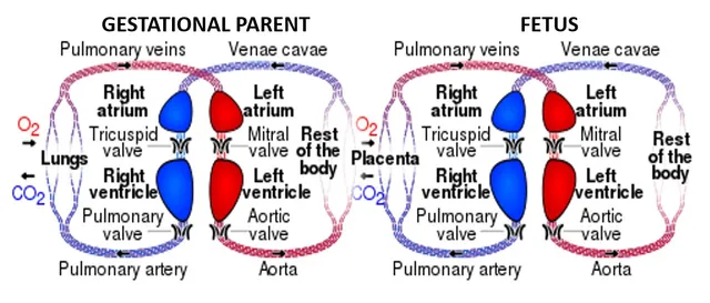
In the fetus, the lungs are collapsed and offer considerable resistance; the placenta, for its part, offers very low resistance. Blood therefore takes three shunts: the ductus venosus bypasses the liver, the foramen ovale passes blood from the right atrium to the left atrium, and the ductus arteriosus connects the pulmonary trunk to the aorta.
At birth, everything reverses within minutes. The first breath unfolds the alveoli and collapses pulmonary resistance; clamping the cord removes the low-resistance placental bed. Pressure becomes higher on the left than on the right, which presses the septum closed and functionally shuts the foramen ovale.
Anatomical closure, however, takes months, and remains incomplete in a notable share of adults — a patent foramen ovale is found in about a quarter of the population.

Amnion, amniotic fluid and the umbilical cord
The amniotic cavity enlarges until it surrounds the whole embryo. Amniotic fluid cushions it against shocks, allows it to move and its limbs to grow symmetrically, and plays a direct part in lung development: the fetus inhales and swallows it.
Its volume reflects the balance between production and removal. In the second half of pregnancy it comes mainly from fetal urine and is removed mainly by swallowing. Oligohydramnios therefore points to a renal or urinary anomaly; polyhydramnios, to an obstacle to swallowing, such as oesophageal atresia.
The umbilical cord links the fetus to the placenta. It normally contains two arteries, which carry deoxygenated fetal blood back to the placenta, and a single vein, which brings oxygenated blood, all embedded in a protective connective tissue, Wharton's jelly.
Building the placenta
After implantation, the trophoblast sends projections into the maternal blood: the villi. They are at first made of trophoblast alone, then receive a core of mesoderm, then fetal vessels; by the end of the third week, the tertiary villi link the embryo's circulation to the pools of maternal blood.
The villi persist only on the side of the uterine wall, where they form the villous chorion, the fetal part of the placenta; on the side facing the uterine cavity they disappear and the chorion becomes smooth. The maternal part of the placenta is the decidua basalis, the transformed uterine lining beneath which the embryo implanted.
The human placenta is called haemochorial: maternal blood directly bathes the trophoblast. By late pregnancy, the barrier between the two bloods thins until, in places, it is reduced to the syncytiotrophoblast and the endothelium of fetal capillaries. At term the placenta weighs about 500 grams, roughly a sixth of the newborn's weight.
Exchanges use several routes. Oxygen and carbon dioxide diffuse; glucose passes through transporters; IgG antibodies are actively transported, which gives the newborn protection during its first months. Fetal haemoglobin has a higher affinity for oxygen than adult haemoglobin, which favours the transfer of oxygen to the fetus.
The hormones of pregnancy
hCG is secreted by the syncytiotrophoblast from implantation onwards. Detectable in the blood about ten days after fertilisation, it is the signal used by pregnancy tests. Its concentration roughly doubles every two days in early pregnancy, peaks around the tenth week, then falls. Its role is to maintain the corpus luteum of the ovary, which secretes the progesterone essential to early pregnancy.
Around the eighth to tenth week, the placenta produces enough progesterone to take over from the corpus luteum. Progesterone maintains the uterine lining and keeps the uterine muscle at rest. For oestrogens, the placenta depends on the fetus: it can make them only from precursors supplied by the fetal adrenal gland, and one speaks of the feto-placental unit.
Human placental lactogen rises throughout pregnancy. It makes the mother less sensitive to insulin, which reserves glucose for the fetus. When the maternal pancreas cannot keep up with this increased demand, gestational diabetes appears, screened for by measuring blood glucose during pregnancy.
When the placenta is diseased
During the first half of pregnancy, trophoblast cells invade the spiral arteries of the uterus and turn them into wide, muscle-free vessels that deliver high flow at low pressure. When this remodelling is inadequate, the placenta is poorly perfused and releases substances that damage the maternal endothelium: this is pre-eclampsia, defined by hypertension and proteinuria after the twentieth week. It threatens mother and child; only delivery cures it. In women at risk, low-dose aspirin started early reduces its frequency.
The hydatidiform mole is an abnormality of fertilisation. In a complete mole, the oocyte has lost its chromosomes and the whole genome comes from the father: there is no embryo, the villi swell into vesicles and hCG reaches very high levels. Monitoring of hCG is needed after evacuation, as a trophoblastic tumour can follow.
The placenta can also implant too low, over or near the cervix, which is placenta praevia and bleeds in the third trimester; or penetrate too deeply into the uterine wall, which is placenta accreta, favoured by caesarean scars and a source of severe haemorrhage at delivery.
The yolk sac and allantois
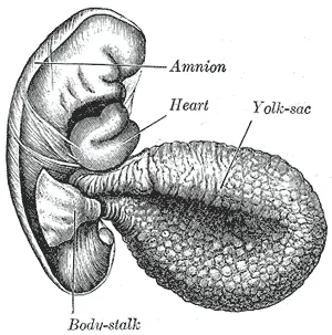
The yolk sac, with no nutrient store in humans, nevertheless plays early roles. It is in its wall that the first blood cells and the first vessels appear, from the third week. It is also where the primordial germ cells are found, the future oogonia or spermatogonia, which then migrate to the developing gonads. The sac is later largely incorporated into the gut during body folding.
The allantois is a small outpouching of the yolk sac that extends into the stalk linking the embryo to the trophoblast. Its vessels become the umbilical vessels. Its intraembryonic part becomes the urachus, a cord linking the apex of the bladder to the umbilicus that persists in adults as a ligament; if it stays open, urine can leak from the newborn's umbilicus.
Prenatal diagnosis
In France, three ultrasound scans are offered during pregnancy. The first-trimester scan, between eleven and fourteen weeks after the last period, dates the pregnancy, counts the embryos and measures the nuchal translucency, the thickness of fluid beneath the skin of the neck, which is greater in trisomy 21. The second- and third-trimester scans look for malformations and monitor growth.
Screening for trisomy 21 combines maternal age, nuchal translucency and markers in maternal blood to calculate a risk. If the risk is high, cell-free fetal DNA circulating in the mother's blood, which actually comes from the placenta, can be analysed; this test is very sensitive but remains a screening test. Only a fetal sample allows diagnosis.
Two sampling methods exist: chorionic villus sampling, possible from eleven weeks, and amniocentesis, sampling of amniotic fluid from fifteen weeks. Both allow the karyotype to be established or a mutation to be sought, at the cost of a small risk of miscarriage.
58 answered questions go with this chapter in the practice section.
Sources and further reading
Reference textbooks
- Gary C. Schoenwolf et coll., Larsen. Embryologie humaine.
Open access
- 28.3 Fetal Development — OpenStax, Anatomy and Physiology 2e.
Last revised : . Text licensed under CC BY-SA 4.0.