Organogenesis and critical periods
Neurulation, germ-layer derivatives, teratogenesis.
Neurulation
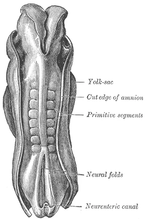
From the third week, the dorsal ectoderm thickens into the neural plate, folds into a groove, then closes into the neural tube. This tube will give the brain at the front and the spinal cord at the back.
A closure defect leaves an open anomaly: anencephaly if the defect is cranial, spina bifida if it is caudal. Folate supplementation before conception markedly reduces this risk.

The critical period
Organogenesis spans the third to the eighth week. It is the period of greatest vulnerability: an insult at this stage causes structural malformations.
Before that, during the first two weeks, the all-or-nothing rule applies: either the embryo dies, or it repairs itself completely. After the eighth week, insults tend to produce functional anomalies or growth restriction.
The neural crest, a de facto fourth germ layer
As the neural tube closes, a population of cells detaches at its junction with the surface ectoderm. These neural crest cells then migrate throughout the embryo, sometimes very far, and give rise to a bewildering variety of derivatives.
They produce the spinal and autonomic ganglia, Schwann cells, the adrenal medulla, melanocytes, and a large part of the facial skeleton. This dispersal makes them the common thread in malformation syndromes that seem unrelated: a migration defect can combine a heart defect, depigmentation and missing colonic ganglia.
Hirschsprung disease is the example: the cells destined for the nerve plexuses of the colon do not complete their migration, and the segment without innervation stays contracted, causing neonatal bowel obstruction.
Heart and gut, two tubes that fold
The heart is the first functional organ: it beats from the end of the third week, since diffusion is already no longer enough. It starts as a single tube, which folds into a loop and then divides into chambers. A septation defect leaves a communication between chambers — ventricular septal defect, the most common congenital heart disease.
The gut faces a space problem. Around the sixth week, the primitive intestinal loop, too long for the abdomen, herniates into the umbilical cord. There it rotates two hundred and seventy degrees counterclockwise, then returns to the abdomen around the tenth week, the cecum settling in the right iliac fossa.
A rotation defect leaves the root of the mesentery abnormally short, and the intestine can then twist around it: this is volvulus, a surgical emergency in infants.
Partitioning of the heart
The primitive heart tube bends into a loop to the right during the fourth week, which places the primitive atrium behind and above the ventricle. Between the fourth and seventh weeks, it is divided into four chambers, and this is when most heart malformations arise, the most common of all congenital malformations.
The atria are separated by two successive partitions. The septum primum grows down from the roof of the atrium; before it closes, an opening forms in its upper part. The thicker septum secundum then grows to its right without ever closing completely. The two offset partitions leave an oblique passage between them, the foramen ovale, which works as a flap valve: blood passes from right to left in the fetus, never the other way.
The ventricle is divided by a muscular septum that rises from the apex, completed above by a small membranous part. This membranous part, the last to close, is the most common site of ventricular septal defects. The single outflow tract, the truncus arteriosus, is divided by a spiral septum into the aorta and pulmonary trunk, which wind around each other.
A defect of this spiral septum explains two major malformations. If it does not spiral, the aorta arises from the right ventricle and the pulmonary artery from the left: this is transposition of the great arteries, incompatible with life without prompt intervention. If it is displaced forwards, it narrows the pulmonary outflow and leaves a ventricular septal defect, over which the aorta sits astride: this is tetralogy of Fallot.
The pharyngeal apparatus
On each side of the future neck, pharyngeal arches form, separated on the outside by clefts and on the inside by pouches. Each arch contains a cartilage core, muscles, an artery and a cranial nerve, and its derivatives remain supplied by that nerve in the adult.
The first arch gives the mandible, two ossicles of the middle ear, the malleus and incus, and the muscles of mastication, supplied by the trigeminal nerve. The second arch gives the stapes, part of the hyoid bone and the muscles of the face, supplied by the facial nerve. The following arches give the rest of the hyoid, the cartilages of the larynx and the muscles of the pharynx and larynx.
The pouches give rise to organs. The first gives the middle ear cavity and the auditory tube. The third gives the thymus and the inferior parathyroids, the fourth the superior parathyroids. The thyroid arises elsewhere, from the floor of the pharynx at the base of the future tongue, and descends into the neck; a remnant of its path can form a cyst in the midline of the neck.
The 22q11 deletion syndrome illustrates this common origin: abnormal development of the third and fourth pouches leads to an absent or small thymus, with immune deficiency, and small parathyroids, with low calcium, together with outflow-tract heart defects and characteristic facial features.
Kidneys and gonads
Three renal systems follow one another, from top to bottom. The pronephros, rudimentary, soon disappears. The mesonephros works briefly then regresses, but its duct, the Wolffian duct, persists. The metanephros, the definitive kidney, arises from the meeting of a bud from the Wolffian duct, the ureteric bud, which will give the ureter, pelvis, calyces and collecting ducts, and a cluster of mesoderm, the blastema, which will give the nephrons.
The definitive kidney arises in the pelvis and ascends to the lumbar region. If the two lower poles fuse, the horseshoe kidney is stopped in its ascent by the inferior mesenteric artery. The fetal kidney produces the urine that forms most of the amniotic fluid: absence of both kidneys leads to a lack of fluid and to failed lung development.
The gonads remain undifferentiated until the seventh week. In an XY embryo, the SRY gene on the Y chromosome steers the gonad towards a testis. The testis secretes anti-Müllerian hormone, which makes the Müllerian ducts regress, and testosterone, which keeps the Wolffian ducts, the future epididymis, vas deferens and seminal vesicles. Dihydrotestosterone, derived from testosterone, masculinises the external genitalia.
Without a testis, the Wolffian ducts regress, the Müllerian ducts persist and form the tubes, uterus and upper vagina, and the external genitalia become female. Complete androgen insensitivity in an XY embryo therefore gives internal testes, no uterus and female external genitalia.
The limbs
The limb buds appear at the end of the fourth week, the upper limb buds slightly before the lower. Each bud is a mass of mesoderm covered by ectoderm, whose rim forms a thickening, the apical ectodermal ridge, which sustains outward growth of the bud: the segments are laid down from root to tip.
A small area of the posterior border of the bud, the zone of polarising activity, secretes a signal that organises the axis from thumb to little finger. Around the sixth week, the tip flattens into a paddle; apoptosis then hollows out the spaces between the fingers. If it fails, the fingers remain joined: this is syndactyly.
Thalidomide, prescribed in the late 1950s for nausea in pregnancy, showed how much timing matters: taken during a narrow window of the second month, it caused severely shortened limbs, with hands or feet attached directly to the trunk.
The main teratogens
A teratogen is an agent that causes a malformation during pregnancy. Its effect depends on the timing of exposure, the dose and the genetic susceptibility of mother and embryo.
Alcohol is the leading avoidable teratogen in rich countries. Fetal alcohol syndrome combines growth restriction, particular facial features and impaired brain development; no dose is recognised as safe. Among drugs, isotretinoin, prescribed for severe acne, causes serious malformations, hence mandatory contraception; valproic acid, an antiepileptic, multiplies the risk of spina bifida and neurodevelopmental disorders; vitamin K antagonists affect the skeleton and nose.
Some infections cross the placenta. Rubella caught in the first trimester can cause cataract, deafness and heart defects, hence vaccination of young women before pregnancy. Cytomegalovirus is the leading infectious cause of childhood deafness. Toxoplasmosis can affect the eye and brain. Poorly controlled maternal diabetes at conception also increases the risk of malformations, notably of the heart.
58 answered questions go with this chapter in the practice section.
Sources and further reading
Reference textbooks
- Gary C. Schoenwolf et coll., Larsen. Embryologie humaine.
Open access
- 28.2 Embryonic Development — OpenStax, Anatomy and Physiology 2e.
Last revised : . Text licensed under CC BY-SA 4.0.