Energy metabolism
Glycolysis, Krebs cycle, respiratory chain and yields.
Three steps in series
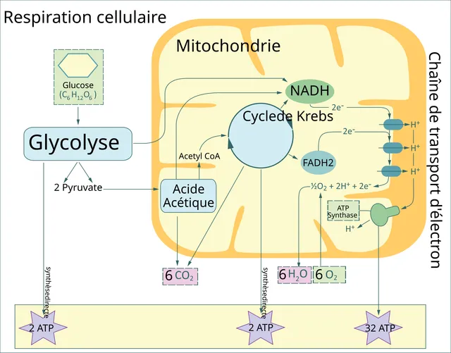
Glycolysis, cytosolic and anaerobic, splits glucose into two pyruvates with a modest net gain. The Krebs cycle, in the mitochondria, completes the oxidation and mainly produces reduced coenzymes. The respiratory chain reoxidizes them and produces most of the ATP.
ATP is therefore not mainly made by the first two steps: their role is to produce NADH and FADH₂, the currency exchanged with the respiratory chain.

Oxidative phosphorylation
Electron transport along the respiratory chain pumps protons out of the matrix and creates a gradient across the inner membrane. The return of these protons through ATP synthase supplies the energy to synthesize ATP.
This is the chemiosmotic theory: energy is not passed chemically from the chain to ATP synthase; it travels through an electrochemical gradient.
The yield, plainly
Glycolysis consumes two ATP and produces four: the net gain is two ATP per glucose, plus two NADH. That is not much, but it works without oxygen — hence its role in red blood cells, which have no mitochondria, and in muscle during intense exertion.
Complete oxidation of pyruvate, on the other hand, delivers most of the energy. Oxidative decarboxylation followed by the Krebs cycle mainly supplies reduced coenzymes, whose reoxidation by the respiratory chain drives ATP synthase. The total approaches about thirty ATP per glucose.
The fifteenfold difference between the two pathways explains how quickly purely anaerobic exertion runs out, and the central place of oxygen in exercise physiology.
Fed, fasting, prolonged fasting
After a meal, insulin dominates: glucose enters muscle and fat cells, the liver builds up its glycogen, fatty acids are stored.
A few hours later, glucagon takes over. The liver breaks down its glycogen and releases glucose — muscle, which lacks glucose-6-phosphatase, keeps its own for itself. This hepatic reserve runs out in about a day.
Beyond that, hepatic gluconeogenesis produces glucose from lactate, glycerol and amino acids, the latter taken from muscle. To limit this wasting, the liver converts fatty acids into ketone bodies, which the brain learns to use within a few days. This adaptation saves several tens of grams of protein a day.
Glycolysis and its control points
Glycolysis converts one molecule of glucose into two pyruvates in the cytosol, in ten reactions. It starts by investing two ATP to phosphorylate glucose and then fructose-6-phosphate; it then recovers four, and produces two NADH. It works in all cells, with or without oxygen.
Three reactions are practically irreversible and serve as control points: those of hexokinase, phosphofructokinase-1 and pyruvate kinase. Phosphofructokinase-1 is the rate-limiting step: it is slowed when the cell is full of energy — abundant ATP and citrate — and stimulated when energy is short — abundant AMP.
Pyruvate then has two fates. With oxygen, it enters the mitochondrion, where pyruvate dehydrogenase converts it into acetyl-CoA. Without oxygen, or in the red cell, which has no mitochondria, it is reduced to lactate: this reaction regenerates the NAD+ without which glycolysis would stop.
The Krebs cycle and the respiratory chain
In the mitochondrial matrix, acetyl-CoA condenses with oxaloacetate to form citrate, which runs through a cycle of reactions and regenerates oxaloacetate. With each turn, two carbons are removed as carbon dioxide, and the cycle produces three NADH, one FADH2 and one GTP, equivalent to one ATP.
The reduced coenzymes give their electrons to the respiratory chain, a series of protein complexes in the inner mitochondrial membrane. The electrons descend towards oxygen, which captures them at the end of the chain and forms water. This transport pumps protons out of the matrix; their return through ATP synthase provides the energy for ATP synthesis.
Cyanide blocks the last complex of the chain: oxygen is present but unusable, and cells die of asphyxia despite well-oxygenated blood. Uncouplers, conversely, let protons return without passing through ATP synthase: the energy is dissipated as heat. This is the physiological role of thermogenin in brown adipose tissue, which keeps newborns warm.
Glycogen, the quick store
Glucose is stored as glycogen, a branched polymer that can be built and broken down quickly. The liver and muscles hold most of it, but for different uses. The liver releases it as glucose into the blood to maintain blood sugar; muscle, lacking glucose-6-phosphatase, cannot do so and uses it for itself.
Synthesis, by glycogen synthase, is stimulated by insulin after meals. Breakdown, by glycogen phosphorylase, is stimulated by glucagon in the liver and by adrenaline in liver and muscle. The two enzymes are regulated in opposite directions by phosphorylation: when one is active, the other is restrained.
Liver stores run out after about a day of fasting. Beyond that, blood sugar depends on gluconeogenesis, which makes glucose from lactate, glycerol and amino acids.
Fats, the reserve fuel
The triglycerides of adipose tissue are by far the largest energy store. During fasting, lipolysis splits them into glycerol and fatty acids, released into the blood; insulin restrains it, the fasting hormones stimulate it. Fatty acids enter the mitochondrion with the help of carnitine and are cut, two carbons at a time, into acetyl-CoA: this is beta-oxidation, which yields a great deal of ATP.
When hepatic acetyl-CoA arrives faster than the Krebs cycle can handle, the liver converts it into ketone bodies — acetoacetate and beta-hydroxybutyrate —, a fuel exported to the heart, muscle and, after a few days of fasting, the brain. In the complete absence of insulin, in type 1 diabetes, this production runs out of control: ketone bodies, which are acids, accumulate and cause ketoacidosis.
Fatty acid synthesis, or lipogenesis, takes place instead in the cytosol, from acetyl-CoA, when intake exceeds needs. It consumes NADPH, supplied largely by the pentose phosphate pathway.
The fate of amino acids
Amino acids are not stored. Those not used to build proteins first lose their amine group, transferred to a keto acid by the transaminases, then released as ammonia. Ammonia, toxic to the brain, is converted into urea by the liver, and urea is excreted by the kidney.
The remaining carbon skeleton feeds energy metabolism. Most amino acids are glucogenic: they can give glucose through gluconeogenesis, which explains why prolonged fasting consumes muscle proteins. A few, such as leucine and lysine, are exclusively ketogenic: they give only acetyl-CoA or ketone bodies.
An enzyme deficiency on one of these pathways can be serious: in phenylketonuria, the absence of one enzyme blocks the conversion of phenylalanine, which accumulates and damages the developing brain. Screening at birth allows a low-phenylalanine diet that prevents this damage.
58 answered questions go with this chapter in the practice section.
Sources and further reading
Reference textbooks
- David L. Nelson, Michael M. Cox, Lehninger. Principes de biochimie.
Open access
- 7.4 Oxidative Phosphorylation — OpenStax, Biology 2e.
- 24.2 Carbohydrate Metabolism — OpenStax, Anatomy and Physiology 2e.
Last revised : . Text licensed under CC BY-SA 4.0.