Proteins: structure and folding
Amino acids, four levels of structure, denaturation.
What drives folding
Folding is dictated by the side chains: hydrophobic residues bury themselves in the core of the protein, polar residues stay exposed to the solvent. Salt bridges, hydrogen bonds and disulfide bridges add to this.
A rise in temperature, an extreme pH or a chaotropic agent breaks these weak interactions: the protein denatures and loses its function, without its primary structure being affected.
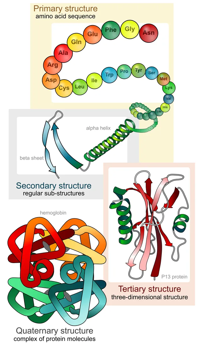
Four levels of organization
The primary structure is the sequence of amino acids, held together by covalent peptide bonds. In principle it contains all the information needed for folding: this is Anfinsen's principle.
The secondary structure describes the regular patterns of the peptide backbone — alpha helix, beta sheet — stabilized by hydrogen bonds between the backbone C=O and N-H groups. Side chains do not take part directly, but they determine a sequence's tendency to form one pattern or the other — proline, for example, breaks helices.
The tertiary structure is the complete three-dimensional fold, this time governed by the side chains: hydrophobic residues bury themselves in the core, polar ones stay exposed, and disulfide bridges can lock the whole thing in place.
The quaternary structure, finally, only exists for proteins with several chains. It is what makes cooperativity possible, hemoglobin being the canonical example.

When folding fails
In the cell, a newly made chain does not always fold on its own. Chaperone proteins, including heat-shock proteins, keep it from aggregating during synthesis and help it reach its conformation; they are overproduced when heat or stress threatens to denature proteins.
A misfolded protein is normally recognised, tagged with ubiquitin and destroyed by the proteasome. When this quality control is overwhelmed, abnormal proteins stack into insoluble fibres rich in beta sheets, amyloid deposits, which build up in tissues.
A single substitution is sometimes enough to change how a protein behaves. In sickle cell disease, the valine that replaces a glutamic acid creates a hydrophobic patch on the surface of deoxygenated haemoglobin: the molecules assemble into long fibres that distort the red cell into a sickle.
Amino acids
Proteins are built from twenty amino acids. Each carries, on a single carbon called alpha, an amine group, a carboxylic acid group, a hydrogen and a side chain of its own. At physiological pH, the amine is protonated and the acid ionised: the amino acid carries both a positive and a negative charge.
Amino acids are classified by their side chain. Some are non-polar, like those of leucine or valine, and bury themselves in the core of proteins. Others are polar and uncharged, like serine. Others still are charged at physiological pH: negatively for aspartate and glutamate, positively for lysine and arginine; histidine, whose side chain has a pKa close to six, can take up or give up a proton depending on the environment.
Nine amino acids cannot be synthesised by adults and must come from the diet: these are the essential amino acids, including leucine, lysine, methionine, phenylalanine and tryptophan. A diet lacking them does not allow proteins to be made, whatever the intake of other amino acids.
The peptide bond
Two amino acids join through a peptide bond, formed between the acid group of one and the amine group of the other, with the loss of a water molecule. A chain of amino acids therefore has a direction: it begins with a free amine end, called the N-terminus, and ends with a free acid end, called the C-terminus. Sequences are always written in this direction, which is also that of translation.
The peptide bond has partial double-bond character: it is planar and does not rotate. The chain's flexibility comes from the two bonds on either side of each alpha carbon, around which rotation is possible but limited by the bulk of the side chains. These constraints explain why a small number of regular conformations — helix, sheet — are found in almost all proteins.
Fibrous and globular proteins
Fibrous proteins are elongated, insoluble and play a mechanical role. Collagen, the most abundant protein in the body, is a triple helix of chains in which every third amino acid is glycine, the only one small enough to fit at the centre of the twist. Its stability requires hydroxylation of proline, which depends on vitamin C: its deficiency, scurvy, makes vessels and gums fragile and prevents wound healing.
Keratin forms hair, nails and the horny layer of the skin; elastin gives arteries and lungs their elastic recoil. Globular proteins, by contrast, are folded into a compact ball and are soluble: enzymes, peptide hormones, antibodies, carriers such as haemoglobin and albumin.
Myoglobin and haemoglobin
Myoglobin, the muscle protein, has only one chain and one haem: it binds oxygen with high affinity along a hyperbolic curve, and serves as a reserve that it releases only when the partial pressure of oxygen becomes very low, during intense exercise.
Adult haemoglobin combines four chains — two alpha and two beta —, each with its haem. Binding one oxygen molecule makes it easier to bind the next: this is cooperativity, which gives the saturation curve its sigmoid shape. Haemoglobin loads almost completely in the lung and unloads extensively in the tissues, over a narrow pressure range.
Several factors lower its affinity and promote oxygen release: acidity and carbon dioxide — the Bohr effect —, heat and 2,3-diphosphoglycerate. Fetal haemoglobin, made of two alpha and two gamma chains, binds the latter less and therefore has a higher affinity, which lets the fetus take oxygen from maternal blood. Carbon monoxide binds haem about two hundred times more strongly than oxygen.
58 answered questions go with this chapter in the practice section.
Sources and further reading
Reference textbooks
- David L. Nelson, Michael M. Cox, Lehninger. Principes de biochimie.
Open access
- 3.4 Proteins — OpenStax, Biology 2e.
Last revised : . Text licensed under CC BY-SA 4.0.