Diffusion and capillary exchange
Fick's law, Starling forces, edema.
Diffusion
Diffusion flux is proportional to the exchange surface area and to the concentration gradient, and inversely proportional to the thickness of the barrier. This is Fick's law.
The alveolar-capillary barrier shows how all three terms are optimized: an immense surface area, a thickness of less than one micrometer, and a gradient maintained by ventilation on one side and perfusion on the other.
The Starling balance
In the capillary, two forces oppose each other. Hydrostatic pressure pushes water out of the vessel; oncotic pressure, due to plasma proteins, holds it in.
The net result pushes fluid out at the arteriolar end and draws it back in at the venous end. The surplus is drained by the lymphatics.
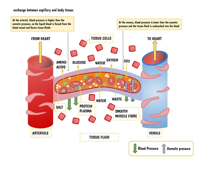

Starling forces in the capillary
Four pressures decide the direction of water movement across the capillary wall. Capillary hydrostatic pressure pushes out, plasma oncotic pressure holds in; their interstitial counterparts act in the opposite direction and are weaker.
At the arteriolar end, hydrostatic pressure wins and there is filtration. It falls along the capillary, so that at the venular end oncotic pressure wins and there is reabsorption. The surplus that is not reabsorbed is taken up by the lymphatics. This is the classic model; we now know that venular reabsorption is limited in the steady state, and that lymphatic drainage accounts for a larger share of the return of filtered water.
Edema reflects a breakdown of this balance, and its mechanism follows from the imbalance involved: high hydrostatic pressure in heart failure, collapsed oncotic pressure in nephrotic syndrome or liver failure, increased permeability in inflammation, interrupted drainage in lymphedema.
Diffusion, fast over short distances only
Diffusion is the spontaneous movement of molecules from a concentrated area to a less concentrated one, driven by their thermal agitation. According to Fick's law, flux is proportional to the diffusion coefficient, the area and the concentration difference, and inversely proportional to the thickness crossed. The diffusion coefficient falls as molecular size increases and as the medium's viscosity increases.
The time needed to diffuse grows as the square of the distance. An oxygen molecule crosses the alveolar wall, about half a micrometre thick, in a fraction of a millisecond; it would take a few seconds for a hundred micrometres, but hours for a centimetre.
This is the reason for the circulation: diffusion suffices over short distances but not beyond. Almost every cell in the body is thus within about a hundred micrometres of a capillary, and poorly vascularised tissues, such as cartilage, stay thin or work at a low rate.
Osmotic pressure and tonicity
When a membrane lets water through but not a solute, water moves to the more concentrated side: this is osmosis. The pressure that would have to be applied to stop it is the osmotic pressure; according to van 't Hoff's law, it is proportional to the total concentration of particles and to absolute temperature. A difference of just one milliosmole per litre corresponds to about 19 mmHg: small concentration differences create considerable forces.
What counts is the number of particles, not their mass. One mole of sodium chloride, which dissociates into two ions, counts as about two osmoles; one mole of glucose as just one.
Tonicity describes a solution's effect on cell volume. Only solutes that do not cross the cell membrane count. Urea, which crosses it freely, raises osmolarity without changing tonicity. Placed in a hypotonic solution, a red cell swells and may burst: this is haemolysis; in a hypertonic solution, it shrinks. 0.9% saline is isotonic with plasma.
Membrane transport
Gases and small lipid-soluble molecules cross the lipid bilayer directly: this is simple diffusion, whose flux is proportional to the gradient without limit. Ions and polar molecules need proteins. Channels form selective pores and are very fast. Carriers bind their solute and change shape to move it across: they are slower and saturable, with a maximum rate reached when all are occupied, like the glucose carriers.
These two modes, called passive, follow the electrochemical gradient and use no energy. Primary active transport goes against the gradient using ATP directly: the sodium-potassium pump moves three sodium ions out and two potassium ions in per molecule of ATP. Secondary active transport uses the sodium gradient created by the pump to drive another solute, in the same direction, like sodium-glucose cotransport in the intestine and kidney, or in the opposite direction, like the exchanger that removes calcium from the cardiac muscle cell.
Large molecules cross in vesicles: endocytosis to enter, exocytosis to leave, transcytosis to pass right through a cell, like IgG across the placenta.
Three kinds of capillaries
The capillary wall is not the same everywhere, and its structure determines what passes. Continuous capillaries, with an uninterrupted endothelium, are found in muscle, skin and lungs; water and small water-soluble molecules pass through the clefts between cells, proteins hardly at all. In the brain, these clefts are closed by tight junctions: this is the blood-brain barrier.
Fenestrated capillaries are perforated by small pores, closed by a thin diaphragm or not. They are found where exchanges of water and small molecules are intense: the renal glomerulus, intestinal villi, endocrine glands, choroid plexuses.
Discontinuous capillaries, or sinusoids, are wide, with large gaps between cells and an incomplete basal lamina. They let proteins and even cells through: they are found in the liver, which releases its proteins into the blood, the bone marrow, from which new blood cells emerge, and the spleen.
Dialysis and ultrafiltration
Haemodialysis replaces part of the kidneys' work by running blood on one side of a semi-permeable membrane and a fluid, the dialysate, on the other, in countercurrent. Small molecules — urea, creatinine, potassium — diffuse from blood to dialysate, where their concentration is zero or low; cells and proteins stay in the blood. The dialysate contains sodium, bicarbonate and calcium at chosen concentrations to correct the patient's balance.
Excess water is removed by ultrafiltration: a pressure difference applied across the membrane pushes water out of the blood, along with the small solutes it carries. Diffusion mainly clears small molecules; ultrafiltration, by convection, clears middle-sized molecules better.
Peritoneal dialysis uses the patient's peritoneum as the membrane: a dialysate is introduced into the abdomen, takes up waste by diffusion, and is then replaced. The glucose it contains, by making it hypertonic, draws water from the blood by osmosis.
58 answered questions go with this chapter in the practice section.
Sources and further reading
Reference textbooks
- John E. Hall, Michael E. Hall, Guyton et Hall. Physiologie médicale, Elsevier Masson.
Open access
- 12.7 Molecular Transport Phenomena: Diffusion, Osmosis, and Related Processes — OpenStax, College Physics 2e.
- 20.3 Capillary Exchange — OpenStax, Anatomy and Physiology 2e.
Last revised : . Text licensed under CC BY-SA 4.0.