Radiation and medical imaging
Ionizing or not, principles of the main modalities, radiation protection.
Ionizing or not
Radiation is called ionizing when it has enough energy to knock an electron out of an atom. X-rays and gamma rays are ionizing; ultrasound, magnetic fields and visible light are not.
This distinction drives all of radiation protection: only ionizing radiation carries a risk of DNA damage, and therefore a delayed cancer risk.
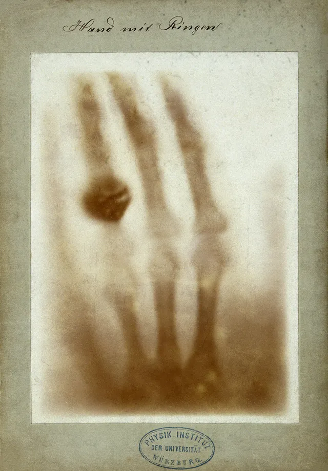

Four principles, four images
Each modality exploits a different physical property, and therefore sees something different.
- Radiography and CT measure the attenuation of X-rays: they mainly see differences in density, which is why they excel at bone.
- Ultrasound measures the reflection of sound waves at interfaces: it sees soft tissues and fluids, but bone and air stop it.
- MRI exploits the resonance of protons in a magnetic field: it offers the best soft-tissue contrast, without ionizing radiation.
- Nuclear medicine injects a radioactive tracer and detects its radiation: it images a function rather than a structure.
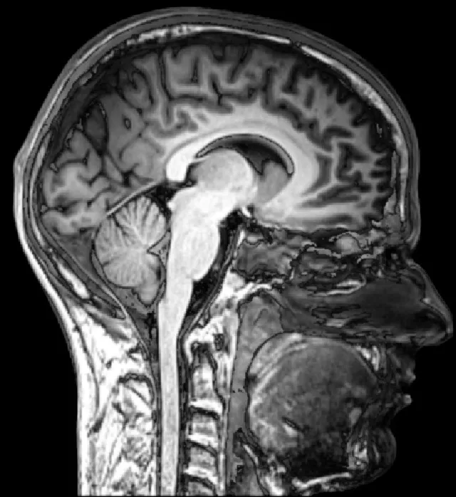

Choosing the examination according to what you are looking for
Radiography and CT rely on the attenuation of X-rays, and therefore on the density and composition of tissues. They excel at bone, calcifications and air, and expose the patient to ionizing radiation — which means every examination must be justified, especially in children and pregnant women.
Magnetic resonance imaging exploits the magnetic properties of protons. It does not irradiate and offers far better contrast in soft tissues: brain, spinal cord, joints. Cortical bone, on the other hand, being poor in mobile protons, stays silent.
Ultrasound relies on the reflection of sound waves at interfaces. It is accessible, portable, repeatable and has no known danger, but air and bone reflect almost everything: the aerated lung and the adult brain remain largely closed to it.
Radioactivity
An unstable nucleus transforms spontaneously by emitting radiation: this is a decay. Alpha decay emits a helium nucleus, highly ionising but stopped by a sheet of paper or the outer layer of the skin. Beta-minus decay emits an electron; beta-plus decay emits a positron, which almost immediately annihilates with an electron, producing two 511 keV photons travelling in opposite directions. The resulting nucleus, often excited, de-excites by emitting a gamma photon.
The activity of a source, in becquerels, is the number of decays per second. It halves with each period, or physical half-life: after n periods, a fraction of 1/2 to the power n remains. After ten periods, about a thousandth of the initial activity remains.
Nuclear medicine chooses its isotopes according to these properties. Technetium 99m, with a six-hour half-life, emits gamma radiation well detected by gamma cameras: it is the isotope of scintigraphy. Fluorine 18, a positron emitter with a half-life of about two hours, is used for positron emission tomography. Iodine 131, a beta-minus emitter with an eight-day half-life, is used to destroy thyroid tissue.
X-rays in matter
An X-ray beam passing through matter is attenuated exponentially: each equal thickness stops the same proportion. The half-value layer is the thickness that halves the intensity; it is much smaller for lead than for tissues, hence its use for aprons and room walls.
Two interactions dominate at diagnostic energies. In the photoelectric effect, the photon is entirely absorbed by an electron of an atom. This effect increases very steeply with atomic number and decreases as energy increases: it creates the contrast between bone, rich in calcium, and soft tissues, and makes iodine and barium so visible. In the Compton effect, the photon gives up part of its energy and is deflected: it dominates in soft tissues, provides no useful contrast, and the scattered radiation it produces is the main source of staff exposure.
Doses and biological effects
Absorbed dose, in grays, measures the energy deposited per kilogram of matter. Effective dose, in sieverts, takes into account the type of radiation and the sensitivity of the organs exposed: it is what allows exposures to be compared. In France, average natural exposure is of the order of three millisieverts per year. A chest X-ray delivers a few hundredths of a millisievert, an abdominal CT scan several millisieverts.
Deterministic effects appear only above a dose threshold, and their severity increases with dose: skin burns, hair loss, cataract, marrow failure. They concern high doses, accidents or radiotherapy. Stochastic effects, cancers and, in theory, heritable abnormalities, have no accepted threshold: it is their probability, not their severity, that increases with dose.
Tissues whose cells divide a lot are the most sensitive: bone marrow, digestive lining, gonads. Children are more sensitive than adults, and embryos even more so, hence the caution with a woman of childbearing age.
Protecting oneself
Radiation protection rests on three principles. Justification: an irradiating examination is done only if its expected benefit exceeds its risk, and if no non-irradiating examination can answer the question. Optimisation: the dose must be as low as reasonably achievable to obtain the information. Limitation: annual limits apply to exposed workers and to the public, but not to patients, for whom the examination is justified case by case.
For staff, three means reduce exposure. Time: dose is proportional to duration. Distance: dose falls as the square of the distance from the source, so doubling the distance divides it by four. Shielding: lead aprons and screens. Exposed workers wear a dosimeter that measures the dose received.
The physics of MRI
MRI uses hydrogen nuclei, very abundant in water and fat. Placed in a strong magnetic field, 1.5 to 3 teslas in routine practice, they align on average with the field. A radiofrequency wave briefly tips them over; as they return to equilibrium, they emit a signal that antennas pick up. The speed of this return, described by two relaxation times, T1 and T2, differs between tissues: this is what creates contrast.
In T1 weighting, fat is bright and water dark; in T2 weighting, water is bright, which highlights oedema, inflammation and many lesions. The MRI contrast agent is based on gadolinium, which shortens the T1 of the tissues where it concentrates.
MRI involves no radiation, but its magnetic field requires precautions. A ferromagnetic object can be violently attracted; a metallic foreign body in the eye can move and damage the retina; some older pacemakers can malfunction. A systematic questionnaire therefore precedes every examination.
58 answered questions go with this chapter in the practice section.
Sources and further reading
Open access
- 32.1 Diagnostics and Medical Imaging — OpenStax, College Physics 2e.
- 32.2 Biological Effects of Ionizing Radiation — OpenStax, College Physics 2e.
Last revised : . Text licensed under CC BY-SA 4.0.