Acid-base equilibria
pH, pKa, buffers and physiological systems.
pH and pKa
pH measures the concentration of protons; pKa characterizes an acid-base pair. When pH equals pKa, the acidic and basic forms are present in equal amounts.
The Henderson-Hasselbalch equation links the two and predicts the ionization state of a molecule at a given pH — a decisive question for drug absorption.

The bicarbonate buffer
The main plasma buffer pairs carbonic acid with the bicarbonate ion. Its effectiveness owes less to its pKa, which is far from blood pH, than to its open nature: CO₂ is eliminated by the lungs and bicarbonate is regulated by the kidneys.
This dual regulation works at two speeds. The lungs correct within minutes by adjusting ventilation; the kidneys take hours or days, adjusting bicarbonate reabsorption and proton excretion.
Reading an acid-base disorder in three questions
Faced with a blood gas, the analysis always follows the same order. First the pH: below 7.38 there is acidemia, above 7.42 alkalemia. Then the origin: if carbon dioxide changes in the direction that explains the pH, the disorder is respiratory; if it is the bicarbonate, it is metabolic.
Finally, compensation. The body never overcompensates: it brings the pH back toward normal without crossing it, and usually without reaching it. A strictly normal pH with both carbon dioxide and bicarbonate abnormal should therefore suggest a mixed disorder, rather than a perfectly compensated simple one.
The timescales differ considerably. Respiratory compensation of a metabolic disorder sets in within minutes, simply by adjusting ventilation. Renal compensation of a respiratory disorder takes hours to days, the time needed to adjust bicarbonate reabsorption.
Acids, bases and pKa
According to Brønsted's definition, an acid is a species able to give up a proton, a base a species able to accept one. An acid and the base it becomes by losing its proton form a pair: carbonic acid and bicarbonate, ammonium and ammonia.
The strength of an acid is measured by its dissociation constant, Ka, or more conveniently by its pKa, the negative logarithm of Ka: the lower the pKa, the stronger the acid. A strong acid, such as hydrochloric acid, is fully dissociated in water; a weak acid, such as acetic acid, only partly.
pH is the negative logarithm of the hydrogen ion concentration. Because the scale is logarithmic, a fall of one pH unit multiplies that concentration by ten. At the normal blood pH of 7.40, it is only about forty nanomoles per litre, a tiny concentration that the body nevertheless defends with great precision.
Calculating a pH, understanding a buffer
For a strong acid, pH follows directly from concentration: a 0.01 mol/L hydrochloric acid solution has a pH of 2. For a weak acid-base pair, the Henderson-Hasselbalch equation links pH to pKa and to the concentration ratio: pH equals pKa plus the logarithm of the base-to-acid ratio.
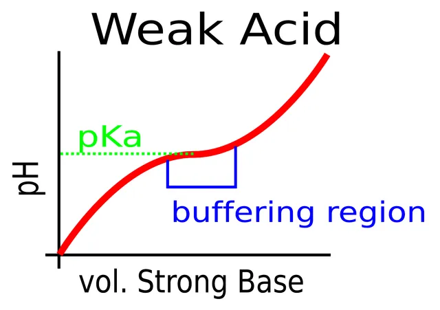
A buffer is a mixture of a weak acid and its conjugate base. When an acid is added, the base takes it up; when a base is added, the acid neutralises it. The pH then changes only slightly. A buffer is most effective when the pH is close to its pKa, within one unit, where both forms are present in comparable amounts.
Applied to blood, the equation is written with the pKa of the bicarbonate pair, 6.1, and the concentration of dissolved carbon dioxide, equal to 0.03 times the PaCO2 in millimetres of mercury. For bicarbonate at 24 mmol/L and a PaCO2 of 40, the ratio is twenty, whose logarithm is 1.3: the pH is 7.4.
The body's buffers
The bicarbonate buffer is the main buffer of extracellular fluid. Its pKa of 6.1 is far from blood pH, but the system is open: the lungs eliminate carbon dioxide and the kidneys adjust the bicarbonate concentration. Each of the two components of the ratio is therefore regulated independently, which makes it an exceptionally effective buffer.
Phosphates, with a pKa around 6.8, buffer mainly inside cells and in urine, where they allow the kidney to excrete hydrogen ions. Proteins, rich in ionisable groups, are the main intracellular buffer; in the red cell, haemoglobin plays a major role, all the more because it takes up protons better once it has released its oxygen.
Buffers only limit the change in pH at the moment. Only the lungs, within minutes, and the kidneys, over days, can actually eliminate the acid load and restore bicarbonate reserves.
The four disorders and their compensations
A disorder is metabolic when it affects bicarbonate first, respiratory when it affects PaCO2 first. The body compensates by changing the other component in the same direction, to bring the ratio, and hence the pH, back towards normal — without ever correcting it completely.
Metabolic acidosis lowers bicarbonate; the lungs compensate by hyperventilating, which lowers PaCO2: this is the deep, sighing Kussmaul breathing of diabetic ketoacidosis. Metabolic alkalosis, for example after heavy vomiting that loses gastric acid, raises bicarbonate; compensation by hypoventilation remains limited, because it runs up against the need for oxygen.
Respiratory acidosis, from hypoventilation, raises PaCO2; the kidney compensates by retaining bicarbonate, but over several days: acute respiratory acidosis is therefore poorly compensated, chronic much more so. Respiratory alkalosis, from hyperventilation — anxiety, altitude, pain —, lowers PaCO2.
The anion gap
The anion gap is calculated as sodium minus the sum of chloride and bicarbonate; it is normally about twelve millimoles per litre. It does not mean that anions are missing: it represents anions that are present but not measured, mainly albumin.
In metabolic acidosis, it points to the cause. A raised gap indicates accumulation of an acid whose anion is not measured: ketone bodies in ketoacidosis, lactate in hypoxia or shock, acids retained in kidney failure, or poisoning by methanol, ethylene glycol or aspirin. A normal gap, with high chloride, points to a loss of bicarbonate, from the gut as in severe diarrhoea, or from the kidney.
58 answered questions go with this chapter in the practice section.
Sources and further reading
Reference textbooks
- Peter Atkins, Loretta Jones, Principes de chimie.
- John E. Hall, Michael E. Hall, Guyton et Hall. Physiologie médicale, Elsevier Masson.
Open access
- 14.6 Buffers — OpenStax, Chemistry 2e.
- 26.4 Acid-Base Balance — OpenStax, Anatomy and Physiology 2e.
Last revised : . Text licensed under CC BY-SA 4.0.