Antibodies and the humoral response
Immunoglobulin structure, isotypes, primary and secondary responses.
One molecule, two functions
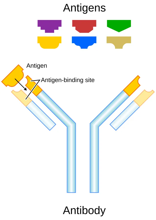
An immunoglobulin pairs two heavy chains with two light chains. Its variable ends carry the antigen-binding sites; its constant fragment determines the effector function — complement activation, opsonization, placental transfer.
The same specificity can therefore be carried by different isotypes: this is class switching, which adapts the function without changing the target.

Primary and secondary
The primary response is slow, of low amplitude, and mainly IgM. The secondary response, on a second contact, is fast, intense and dominated by IgG of much higher affinity.
This difference is the whole principle of vaccination: triggering a primary response without disease, so that real contact immediately sets off a secondary response.
One specificity, five functions
An antibody divides into two functional parts. The Fab fragments carry the antigen-binding sites, generated by rearrangement of the V, D and J segments. The Fc fragment determines the class, and therefore what the antibody triggers once bound.
Class switching keeps the specificity while changing the Fc. A clone that was producing pentameric IgM can thus switch to IgG, IgA or IgE depending on the signals it receives, without changing its recognition at all.
The choice matters. IgG crosses the placenta and activates complement; IgA dimerizes, reaches the secretions and neutralizes without causing inflammation; IgE binds to mast cells and triggers degranulation.
- IgM: produced first, pentameric, strong complement activation, does not cross the placenta.
- IgG: the most abundant, secondary response, crosses the placenta.
- IgA: secretions and breast milk, dimeric in secretions, protected by its secretory component.
- IgE: immediate allergy and defense against parasites, the least abundant.
- IgD: mostly membrane-bound on naive B cells, role still debated.
The germinal centre, workshop of good antibodies
After activation, some B lymphocytes move to the lymph node follicles and form germinal centres there. They multiply very quickly, and an enzyme specific to these cells introduces mutations into their receptor genes: this is somatic hypermutation. Most mutations improve nothing; some increase affinity for the antigen.
The mutated B lymphocytes then compete to capture antigen and obtain help from follicular helper T cells: only those that bind it best survive. Over the weeks, the average affinity of antibodies increases: this is affinity maturation. At the same time, through contact with the T lymphocyte via CD40 and its ligand, and under the influence of cytokines, the B lymphocyte switches antibody class, from IgM to IgG, IgA or IgE, without changing specificity.
The germinal centre produces two kinds of long-lasting cells: long-lived plasma cells, which settle in the bone marrow and secrete antibodies for years, and memory B lymphocytes, ready to respond quickly to a new encounter. When the CD40 ligand is missing, an X-linked disease, class switching fails: the patient has plenty of IgM but almost no IgG, IgA or IgE.
Antigens with and without T lymphocytes
Protein antigens are called thymus-dependent: the B lymphocyte response requires help from CD4 T lymphocytes that recognise peptides from the same protein. It leads to high-affinity antibodies of all classes and to memory.
The capsular polysaccharides of some bacteria, made of one repeated unit, can activate B lymphocytes directly by bringing many receptors together at once. These thymus-independent antigens give mainly IgM, little memory, and a very weak response before the age of two, when this type of response is still immature.
Conjugate vaccines get around this limitation: the polysaccharide is attached to a carrier protein. B lymphocytes that recognise the sugar present peptides of the protein to T lymphocytes, which help them. The response becomes thymus-dependent, with memory, and effective from the first months. This is the principle of infant vaccines against Haemophilus, pneumococcus and meningococcus.
What antibodies do
An antibody kills nothing by itself: it tags and blocks. It neutralises toxins and viruses by masking the sites through which they attach to cells: this is how tetanus and diphtheria vaccines protect. Bound to a microbe, it opsonises it: phagocytes recognise it through receptors for the Fc portion. It activates the classical complement pathway, IgM being the most effective thanks to its five units. Finally, NK cells kill cells coated with IgG: this is antibody-dependent cellular cytotoxicity.
IgG is the most abundant in the blood; it has a half-life of about three weeks, extended by a receptor that recycles it, and the same receptor carries it across the placenta. Secretory IgA protects mucous membranes and passes into breast milk. IgE, very scarce, is bound to mast cells, ready to trigger the immediate reaction.
The four types of hypersensitivity
Type I hypersensitivity, immediate, is allergy: the allergen binds to IgE on mast cells, which release histamine and mediators within minutes. Its generalised form, anaphylaxis, combines a fall in blood pressure, swelling and breathing difficulty; adrenaline injected into muscle is the emergency treatment.
In type II, antibodies bind to antigens on cells or tissues and destroy them, through complement or phagocytes: this is the mechanism of ABO transfusion reactions or some autoimmune anaemias. In type III, circulating antigen-antibody complexes are deposited in vessels, kidneys or joints and trigger inflammation there: serum sickness, some vasculitides, lupus.
Type IV, delayed, involves no antibodies but T lymphocytes. It appears 48 to 72 hours after contact: this is contact eczema, for example to nickel, and the skin reaction to tuberculin, which reflects previous immunity against tuberculosis.
Blood groups and transfusion
Red cells carry the antigens of the ABO system: A, B, both, or none for group O. Each person naturally has, from early childhood, IgM antibodies against the antigens they lack: a group A person has anti-B, a group O person anti-A and anti-B, a group AB person neither. Transfusing incompatible red cells triggers abrupt destruction of the transfused cells, which can be fatal; hence the bedside checks.
For red cells, group O is the universal donor, since it carries neither A nor B, and group AB the universal recipient. For plasma, which contains the antibodies, it is the reverse: AB plasma, with no antibodies, can be given to everyone.
The Rhesus system works differently: a Rhesus-negative person has no natural anti-D antibodies but can make them, of IgG type, after contact with Rhesus-positive red cells. In a Rhesus-negative woman pregnant with a Rhesus-positive child, this contact occurs mainly at delivery; in a later pregnancy, anti-D IgG crosses the placenta and destroys the red cells of a Rhesus-positive fetus. It is prevented by giving the mother anti-D immunoglobulin after each at-risk event, which removes fetal red cells before she becomes immunised.
58 answered questions go with this chapter in the practice section.
Sources and further reading
Reference textbooks
- Kenneth Murphy, Casey Weaver, Janeway. Immunobiologie.
Open access
- 21.4 The Adaptive Immune Response: B-lymphocytes and Antibodies — OpenStax, Anatomy and Physiology 2e.
Last revised : . Text licensed under CC BY-SA 4.0.