Lymphocytes and antigen presentation
T cells, B cells, MHC, selection and tolerance.
Two lineages, two ways of seeing
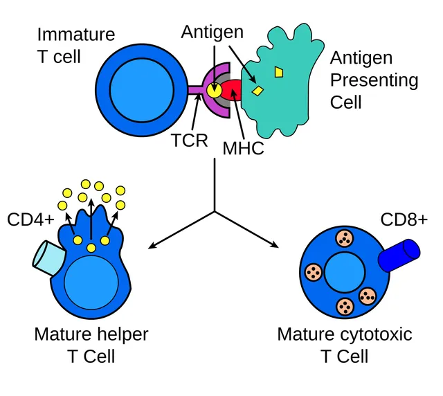
The B cell recognizes the native antigen directly, through its surface immunoglobulin. The T cell only recognizes it when presented, as a peptide, by a major histocompatibility complex molecule.
Selection and tolerance
The lymphocyte repertoire is generated at random by recombination of the receptor genes. It therefore inevitably produces clones directed against self.
Thymic selection eliminates these clones: positive selection of those that recognize MHC, negative selection of those that recognize self too strongly. Peripheral mechanisms complete this tolerance; when they fail, the way opens to autoimmunity.
Presenting antigen: two routes, two audiences
Major histocompatibility complex class I molecules are present on every nucleated cell and present peptides of intracellular origin. They therefore constantly display a sample of what is being made inside — including viral proteins. CD8 T cells monitor them and kill whatever is abnormal.
Class II molecules are expressed mainly by professional antigen-presenting cells and present peptides of extracellular origin, captured and then broken down. CD4 T cells read them and set the coordination of the response in motion.
This division explains the logic of defense: what is outside is handled by antibodies and phagocytosis, under the direction of CD4 cells; what is inside is mainly eliminated by killing the infected cell, the job of CD8 cells.

Making millions of different receptors
The genome does not contain one gene per receptor: it contains gene segments that each maturing lymphocyte assembles at random, one segment of each type. This rearrangement, called VDJ, is carried out by enzymes, the RAG recombinases. At the junctions, nucleotides are also removed or added at random, which multiplies diversity further. The number of possible receptors far exceeds the number of lymphocytes in an individual.
Each lymphocyte carries a single type of receptor, and therefore a single specificity. When an antigen meets the lymphocyte whose receptor matches it, that lymphocyte alone is selected and multiplies: this is clonal selection, followed by clonal expansion that produces thousands of identical cells.
Without recombinases, no functional B or T lymphocyte can form: this is one of the causes of severe combined immunodeficiency, in which the infant has serious infections from the first months. Haematopoietic stem cell transplantation is the treatment.
The MHC, or HLA system
In humans, the molecules of the major histocompatibility complex are called HLA; their genes are clustered on chromosome 6. Class I molecules, HLA-A, B and C, are present on almost all nucleated cells; they present peptides derived from cytoplasmic proteins, cut up by the proteasome and brought into the reticulum by a transporter. CD8 T lymphocytes read them: any virus-infected cell can thus be spotted.
Class II molecules, HLA-DR, DQ and DP, are present mainly on antigen-presenting cells: dendritic cells, macrophages, B lymphocytes. They present peptides from proteins taken up from outside and degraded in endosomes, and are read by CD4 T lymphocytes.
HLA genes are the most variable in the genome, and both alleles of each gene are expressed together. Two unrelated people are therefore very unlikely to be identical, which complicates transplantation: a donor is first sought among siblings, where a brother or sister has a one-in-four chance of being identical. Some alleles are associated with diseases, such as HLA-B27 with ankylosing spondylitis.
Activating a T lymphocyte, and restraining it
A naive T lymphocyte is activated only if it receives several signals at once. The first is recognition, by its receptor, of a peptide presented by an HLA molecule. The second is a costimulatory signal, for example binding of its CD28 molecule to molecules that the dendritic cell expresses only once activated by danger. Cytokines then direct its differentiation. A lymphocyte that receives the first signal without the second becomes unresponsive: this is anergy, one of the mechanisms of tolerance.
Once activated, the lymphocyte produces interleukin 2, which makes it proliferate. Ciclosporin and tacrolimus, used after transplantation, block this production and prevent rejection.
Other receptors instead restrain activation, such as CTLA-4 and PD-1: these are checkpoints that prevent an excessive response. Many tumours exploit them to protect themselves. Antibodies that block these brakes release the antitumour response; they have transformed the treatment of melanoma and some lung cancers, at the cost of autoimmune side effects.
The specialities of T lymphocytes
CD4 T lymphocytes differentiate into subsets according to the cytokines present at activation. Th1 cells produce interferon gamma, which activates macrophages against intracellular microbes, such as the tuberculosis mycobacterium. Th2 cells produce interleukins that stimulate eosinophils and IgE production: they fight parasites and are involved in allergy. Th17 cells recruit neutrophils against certain bacteria and fungi. Follicular helper T cells help B lymphocytes in germinal centres.
Regulatory T lymphocytes, which express the transcription factor FOXP3, restrain other lymphocytes and maintain tolerance. An inherited defect of FOXP3 causes, from the first months, autoimmunity affecting many organs.
Cytotoxic CD8 T lymphocytes kill cells that present a foreign peptide on their class I molecules. They release perforin, which pierces the target's membrane, and granzymes, which trigger its apoptosis, or they activate the Fas death receptor.
Where lymphocytes live and circulate
Lymphocytes arise in the bone marrow. B cells complete their maturation there; T cells finish theirs in the thymus, very active in childhood and gradually involuting from puberty onwards. These are the primary lymphoid organs.
Secondary lymphoid organs are the meeting places with antigen. In a lymph node, B lymphocytes group in the follicles of the cortex, T lymphocytes in the deeper paracortical zone; the lymph brings antigens and dendritic cells. The spleen filters the blood; its lymphoid areas, the white pulp, respond to circulating antigens. Mucous membranes have their own lymphoid tissue, such as the Peyer's patches of the intestine.
Naive lymphocytes recirculate constantly: they leave the blood through special venules of the lymph nodes, travel through the node, leave through the lymph and return to the blood. Each lymphocyte thus visits many lymph nodes and increases its chances of meeting its antigen. Without a spleen, after removal, the risk of severe infection by encapsulated bacteria such as pneumococcus rises: specific vaccinations are essential.
58 answered questions go with this chapter in the practice section.
Sources and further reading
Reference textbooks
- Kenneth Murphy, Casey Weaver, Janeway. Immunobiologie.
Open access
- 21.3 The Adaptive Immune Response: T lymphocytes and Their Functional Types — OpenStax, Anatomy and Physiology 2e.
Last revised : . Text licensed under CC BY-SA 4.0.