Innate and adaptive immunity
Speed, specificity, memory and cooperation.
Barriers and inflammation
The first line of defense is not cellular: skin, mucous membranes, mucus, acidic pH, commensal flora. An infection first requires these barriers to be breached.
Once they are, local inflammation sets in: vasodilation, increased permeability, recruitment of phagocytes. Redness, heat, swelling and pain are its direct clinical signs.
Two immunities, and the handover from one to the other
Innate immunity is operational within minutes. It recognizes molecular patterns shared by whole families of microorganisms — bacterial walls, viral double-stranded RNA — through receptors encoded in the genome, identical in every individual.
Adaptive immunity takes days to set up. It recognizes a precise antigen, through receptors generated by gene rearrangement and therefore unique to each lymphocyte clone. It keeps a memory.
The two are not simply side by side: the first commands the second. A dendritic cell that has engulfed a pathogen migrates to the lymph node, presents the antigen there and provides the costimulatory signals without which the T cell would not be activated. This explains the role of vaccine adjuvants, whose essential role is to alert innate immunity.

Inflammation, from signal to repair
Mediators released at the site of injury cause vasodilation and increased vascular permeability. Hence the redness, heat and swelling; the pain comes from tissue tension and the sensitization of pain nerve endings.
These changes have a purpose: bringing more blood to the site, and therefore more cells and more plasma proteins. Neutrophils arrive first, within hours, and phagocytose. Monocytes follow, differentiate into macrophages and take over for several days.
Resolution is as active as onset. Macrophages clear the debris and secrete repair factors. When this phase fails, inflammation becomes chronic and itself destructive — the common mechanism of many chronic diseases.
The cells of innate immunity
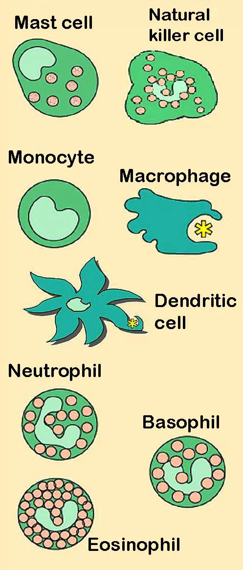
The neutrophil granulocyte is the front-line soldier: very numerous in the blood, it reaches the site of a bacterial infection within hours, phagocytoses and dies there. The macrophage, permanently present in the tissues, also phagocytoses, lives long, releases cytokines that orchestrate inflammation and clears debris. The eosinophil targets parasites too large to be phagocytosed; the mast cell triggers the immediate reaction.
The dendritic cell provides the link to adaptive immunity. A sentinel of the tissues, it captures antigens, then, activated by a danger signal, migrates through the lymph to the lymph node, where it presents these antigens to naive T lymphocytes. Without it, there is no primary adaptive response.
NK cells, lymphocytes of innate immunity, kill virus-infected or tumour cells, notably those that have lost their MHC class I molecules to escape T lymphocytes. They do so without prior activation, by releasing perforin and granzymes.
Recognising danger
Innate immunity does not recognise a precise antigen, but molecular patterns shared by large families of microbes and absent from our cells: lipopolysaccharide from the wall of Gram-negative bacteria, flagellin, the double-stranded RNA of many viruses, certain bacterial DNA sequences. These patterns are detected by receptors encoded in the genome, present identically on all cells of the same type.
Toll-like receptors, on the cell surface or in endosomes, are the best known; other receptors monitor the cytoplasm. The same receptors also detect danger signals released by our own cells when they die abruptly, which explains why trauma or a burn triggers inflammation without any microbe.
Activation of these receptors causes production of inflammatory cytokines — interleukin 1, interleukin 6, TNF — and interferons in the case of viruses. Some, such as interleukin 1, additionally require assembly of a cytoplasmic complex, the inflammasome, which cuts their precursor into the active form.
Complement in three pathways
Complement is a set of about thirty plasma proteins, made mainly by the liver, that activate in a cascade. Three pathways lead to it. The classical pathway starts when antibodies bound to a microbe recruit the protein C1: it therefore links adaptive immunity to complement. The lectin pathway starts when a plasma protein recognises sugars on the microbial surface. The alternative pathway starts spontaneously, at a low level, on any surface that does not restrain it.
The three pathways converge on cleavage of C3. The C3b fragment is deposited on the microbe and makes it recognisable by phagocytes: this is opsonisation, the most important function of complement. The small fragments C3a and C5a attract phagocytes and increase vessel permeability. At the end of the chain, the terminal components, C5b to C9, pierce the membrane of certain bacteria: this is the membrane attack complex.
Our cells carry proteins that restrain complement on their surface. A defect in the C1 inhibitor causes hereditary angioedema: sudden swelling of the skin, gut and larynx, without hives or itching, which can cause suffocation and does not respond to antihistamines.
From vessel to phagolysosome
To reach the site of infection, the neutrophil must leave the vessel: this is diapedesis. It first rolls along the activated endothelium, slowed by selectins; it then stops there, firmly attached by its integrins; finally it squeezes between two endothelial cells. It then climbs the gradient of chemotactic substances up to the microbe.
The phagocyte engulfs the microbe in a vesicle, the phagosome, which fuses with lysosomes and granules: in the phagolysosome, the microbe is attacked by enzymes, antimicrobial peptides and, above all, toxic oxygen derivatives produced by a membrane enzyme, NADPH oxidase: this is the oxidative burst.
Inherited deficiencies illustrate each step. When integrins are missing, neutrophils, unable to leave the vessels, accumulate in the blood and infections occur without pus. When NADPH oxidase is missing, in chronic granulomatous disease, phagocytes swallow microbes without being able to kill them: severe bacterial and fungal infections, and granulomas.
Measuring inflammation, and when it becomes chronic
Interleukin 6 causes the liver to produce acute-phase proteins, including C-reactive protein, or CRP, and fibrinogen. CRP rises within hours and falls quickly when inflammation subsides: it is the marker most used to follow an infection. The erythrocyte sedimentation rate, slower to change, mainly reflects fibrinogen and immunoglobulins. Procalcitonin rises mainly in bacterial infections, which helps decide on antibiotic treatment.
When the injury persists — a microbe hard to eliminate, a foreign body, an autoimmune reaction — inflammation becomes chronic. Neutrophils give way to macrophages and lymphocytes, and destruction goes hand in hand with attempts at repair, with fibrosis that can impair the organ.
The granuloma is a special form of chronic inflammation: macrophages gather in clusters, take on an epithelioid appearance and sometimes fuse into giant cells, to wall off an agent they cannot destroy. In tuberculosis, the centre of the granuloma necroses into a whitish substance, caseous necrosis, which is highly suggestive.
58 answered questions go with this chapter in the practice section.
Sources and further reading
Reference textbooks
- Kenneth Murphy, Casey Weaver, Janeway. Immunobiologie.
Open access
- 21.2 Barrier Defenses and the Innate Immune Response — OpenStax, Anatomy and Physiology 2e.
Last revised : . Text licensed under CC BY-SA 4.0.