Cell cycle and division
Interphase, mitosis, checkpoints and apoptosis.
The four phases
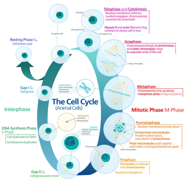
The cycle alternates a long interphase with a brief mitosis. Interphase breaks down into G1, growth; S, DNA replication; G2, preparation for division.
A cell that stops proliferating leaves the cycle in G0. Some stay there for good — neurons, cardiomyocytes; others can be recalled, like hepatocytes after a partial hepatectomy.

Checkpoints
The cycle is punctuated by checkpoints that verify the previous step is complete and error-free. The G1/S checkpoint checks DNA integrity before replicating it; the G2/M checkpoint, before separating it; the spindle checkpoint checks that every chromosome is attached.
The p53 protein is the central player in the G1/S checkpoint. It halts the cycle while repair takes place and, if the damage cannot be repaired, triggers apoptosis.
The cycle and its brakes
The cell cycle is driven by complexes made of a cyclin and a cyclin-dependent kinase. The cyclin is produced and then destroyed at every turn, which gives the system its rhythm; the kinase, for its part, remains present but is only active when paired.
Three checkpoints make sure nothing is set in motion wrongly. The G1 checkpoint examines DNA integrity and the availability of resources before allowing replication. The G2 checkpoint checks that replication is complete and correct. The metaphase checkpoint waits until every chromosome is attached to the spindle by both of its kinetochores.
The p53 protein guards the first one. Faced with DNA damage, it halts the cycle while repair takes place; if the damage is too great, it triggers apoptosis. Its inactivation, present in about half of human cancers, lets the cell replicate with a damaged genome.
Mitosis, step by step
In prophase, chromatin condenses into visible chromosomes, each made of two sister chromatids joined at the centromere; the nucleolus disappears and the two centrosomes move apart to organise the spindle. In prometaphase, the nuclear envelope breaks up and spindle microtubules attach to the kinetochores, protein structures of the centromere.
In metaphase, chromosomes line up on the equatorial plate, each sister chromatid facing a pole. As long as a single kinetochore remains unattached, the spindle checkpoint blocks progress. When all are attached, an enzyme complex triggers the destruction of the protein that restrained the separating enzyme: the cohesins joining sister chromatids are cut all at once.
In anaphase, sister chromatids separate and move to opposite poles. In telophase, a nuclear envelope re-forms around each set and the chromosomes decondense. Cytokinesis, through a contractile ring of actin and myosin, finally divides the cytoplasm.
The engines of the cycle: cyclins and kinases
Progression from one phase to the next is driven by kinases, the CDKs, which are active only when bound to a cyclin. CDKs are present all the time; it is the cyclins, made and then destroyed at precise moments, that set the rhythm. Each cyclin-CDK pair phosphorylates the proteins that trigger the next step.
At the end of G1, cyclin-CDK complexes phosphorylate the retinoblastoma protein, Rb. Unphosphorylated, Rb holds back a transcription factor essential for entry into S phase; phosphorylated, it releases it, and the cell commits to replication. This is the restriction point, beyond which the cell no longer needs growth factors to complete its cycle.
CDK inhibitors slow the cycle. One of them is produced under the influence of p53 when DNA is damaged: the cell stops while repairs are made. Drugs that inhibit certain CDKs are now used against some breast cancers, to block tumour proliferation in G1.
Apoptosis, a programmed death
Apoptosis is an orderly cell suicide, carried out by proteases, the caspases. Two pathways lead to it. The intrinsic, or mitochondrial, pathway responds to internal stress — damaged DNA, lack of growth factors: the outer mitochondrial membrane becomes permeable, cytochrome c is released and activates the caspases. Proteins of the Bcl-2 family regulate this step, some protecting the cell, others pushing it to die.
The extrinsic pathway responds to a signal from outside: binding of a ligand to a death receptor, such as Fas, directly activates an initiator caspase. It is one of the weapons of cytotoxic T cells.
The apoptotic cell shrinks, fragments its DNA and breaks up into membrane-bound apoptotic bodies, which are immediately phagocytosed: there is no leakage of contents and no inflammation. Apoptosis sculpts organs during development — it hollows out the spaces between the fingers — and eliminates self-reactive lymphocytes. Too little apoptosis promotes cancer; too much, neurodegenerative diseases.
The cell cycle and cancer
Cancer arises from the accumulation of mutations in two categories of genes. Proto-oncogenes normally stimulate proliferation; a mutation that makes them overactive turns them into oncogenes, and only one of the two alleles needs to be affected. Tumour suppressor genes, such as RB or TP53, restrain proliferation or trigger apoptosis; in principle both alleles must be lost for the brake to disappear.
Retinoblastoma, a childhood tumour of the retina, illustrated this logic. In the inherited form, the child is born with one RB allele already mutated in every cell: a single second mutation, which is common, is enough for a tumour to appear, often in both eyes and early. In the sporadic form, two mutations are needed in the same cell: the tumour is single and appears later. This is the two-hit hypothesis.
Normal cells have a limited number of divisions, because their telomeres shorten with each replication. Most cancer cells reactivate telomerase, which lengthens them, and become able to divide indefinitely.
Tissues that renew and tissues that do not
Labile cells divide constantly: epidermis, intestinal epithelium, bone marrow. These tissues regenerate quickly, but are also the most affected by treatments that block division, hence hair loss, mouth ulcers and falling blood counts during chemotherapy.
Stable cells, such as hepatocytes, rest in G0 but can re-enter the cycle when the tissue is injured: after part of the liver is removed, the remainder regenerates within weeks. Permanent cells, such as neurons and cardiac muscle cells, have left the cycle: their loss, in a heart attack or stroke, is replaced by a scar rather than by functional tissue.
58 answered questions go with this chapter in the practice section.
Sources and further reading
Reference textbooks
- Bruce Alberts et coll., Biologie moléculaire de la cellule.
Open access
- 10.2 The Cell Cycle — OpenStax, Biology 2e.
- 10.3 Control of the Cell Cycle — OpenStax, Biology 2e.
Last revised : . Text licensed under CC BY-SA 4.0.