Membrane transport
Diffusion, facilitated transport, pumps and vesicles.
A selective barrier
The lipid bilayer spontaneously lets through small nonpolar molecules and, more slowly, water. Everything else — ions, sugars, amino acids — needs a protein to cross.
This selectivity is what makes it possible to maintain very different ionic compositions on either side of the membrane, and therefore for a resting potential to exist.

Passive versus active
Passive transport follows the electrochemical gradient and costs no energy: simple diffusion, and facilitated diffusion through a channel or a carrier.
Active transport moves against the gradient and consumes energy, directly as ATP for primary active transport, indirectly for secondary active transport, which exploits a pre-existing gradient.
Bulk transport
Beyond a certain size, no protein is enough: the cell deforms its membrane. Endocytosis brings material in; exocytosis releases it.
Phagocytosis handles large particles, pinocytosis fluids, receptor-mediated endocytosis specific molecules — this is how LDL cholesterol or iron carried by transferrin gets in.
A bilayer, and why it holds together on its own
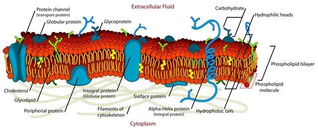
A phospholipid has a polar head, which likes water, and two fatty acid chains that avoid it. Dropped into water, it cannot satisfy both halves at once — except by joining its neighbors, heads facing outward and tails tucked away. The bilayer therefore forms spontaneously, with no enzyme and no energy cost.
This detail has an underestimated consequence: a punctured membrane seals itself. The assembly is not held together by covalent bonds but by water's tendency to organize itself, and that tendency is always at work. This is what makes vesicle fusion, budding and cell division possible.
The fluidity of the whole depends on the length and saturation of the chains: the shorter and more unsaturated they are, the more fluid the membrane. Cholesterol, inserted between the phospholipids, acts as a buffer — it hinders movement when it is warm and prevents stiffening when it is cold.
What gets through, and what does not
The hydrophobic core of the bilayer is a filter by nature. Gases — oxygen, carbon dioxide — and small nonpolar molecules cross it freely. Water, tiny but polar, crosses it slowly. Ions and charged molecules practically do not cross it at all.
All of physiology follows from this selective impermeability. A cell that let ions through freely could maintain no gradient, hence no membrane potential, hence no nerve impulse. The barrier is what makes excitability possible.
For what cannot get through, the cell has dedicated proteins: channels, which open a pore and let substances flow down their gradient; carriers, which change conformation and convey one or a few molecules per cycle; pumps, which spend ATP to work against the gradient.
- Simple diffusion: no protein, no energy, down the gradient.
- Facilitated diffusion: with a protein, no energy, down the gradient — and saturable, since the number of carriers is limited.
- Primary active transport: with a protein, with ATP, against the gradient.
- Secondary active transport: no direct ATP, but exploiting a gradient created by primary transport.
Membrane proteins
Membrane proteins carry out most of the membrane's functions. Integral proteins cross it, often through one or more helices made of hydrophobic amino acids, in contact with the fatty chains of the phospholipids. Extrinsic, or peripheral, proteins simply sit on one face and detach easily. Others still are anchored by a lipid inserted into the bilayer.
These proteins transport, receive signals, catalyse reactions or attach the cell to its neighbours and to the matrix. Cadherins, which adhere only in the presence of calcium, join cells to one another; integrins attach them to the extracellular matrix and pass mechanical tension to the cytoskeleton.
On the outer face, the sugars carried by glycoproteins and glycolipids form the glycocalyx, which acts as an identity signature. Blood groups A, B and O, for example, correspond to variations in these sugar chains on the surface of red cells.
Channels, carriers and pumps
Channels form a water-filled pore that lets a specific ion pass down its gradient at a considerable rate. They open in response to a ligand binding, a change in membrane potential or mechanical stress; they are what produce the electrical signals of neurons and muscles.
Carriers, by contrast, bind their substrate and change shape to move it across: they are slower and saturable, like enzymes. A uniporter moves a single molecule, like the GLUT glucose transporter; a symporter moves two in the same direction, like the intestinal sodium-glucose cotransporter; an antiporter exchanges them in opposite directions, like the sodium-calcium exchanger of the heart.
Pumps use ATP directly to transport against the gradient. Besides the sodium-potassium pump, there are the calcium pumps of the reticulum, the proton pumps of lysosomes, and the large family of ABC transporters, which expel many molecules — including anticancer drugs, which makes some tumours resistant.
Cystic fibrosis, a transport disease
Cystic fibrosis is the most common serious genetic disease in populations of European origin. It is caused by mutations in the CFTR gene, which encodes a chloride channel of the ABC transporter family, located at the apical pole of epithelial cells. The most common mutation removes a single amino acid: the misfolded protein is destroyed before reaching the membrane.
Without chloride leaving the cell, water no longer follows: secretions of the bronchi, pancreas and bile ducts become thick and block the ducts. The bronchi become chronically infected, and the pancreas stops delivering its enzymes, causing poor digestion of fats. In the sweat gland, conversely, chloride is no longer reabsorbed: sweat is abnormally salty, which is the basis of the diagnostic sweat test.
The disease is screened for at birth in France. Drugs that help the mutated CFTR protein fold and open have transformed the outlook for many patients.
Receiving a signal
Hydrophilic molecules, which do not cross the membrane, act through surface receptors. Receptor channels open as soon as the ligand binds, within milliseconds: this is the case of the nicotinic acetylcholine receptor. G-protein-coupled receptors activate a G protein, which in turn modulates an enzyme producing a second messenger.
Two pathways are classic. The Gs protein activates adenylate cyclase, which produces cyclic AMP; this activates a protein kinase that phosphorylates target proteins. The Gq protein activates a phospholipase that releases two messengers, one releasing calcium from the reticulum, the other activating another kinase. Each step amplifies the signal: a single hormone molecule can lead to thousands of second-messenger molecules.
Other receptors carry an enzymatic activity themselves: receptor tyrosine kinases, like the receptor for insulin or for many growth factors, phosphorylate themselves when the ligand binds and recruit signalling proteins.
58 answered questions go with this chapter in the practice section.
Sources and further reading
Reference textbooks
- Bruce Alberts et coll., Biologie moléculaire de la cellule.
Open access
- 5.1 Components and Structure — OpenStax, Biology 2e.
- 5.2 Passive Transport — OpenStax, Biology 2e.
- 5.3 Active Transport — OpenStax, Biology 2e.
Last revised : . Text licensed under CC BY-SA 4.0.