Organelles and protein trafficking
Reticulum, Golgi, targeting and secretory pathways.
Sorting starts at translation
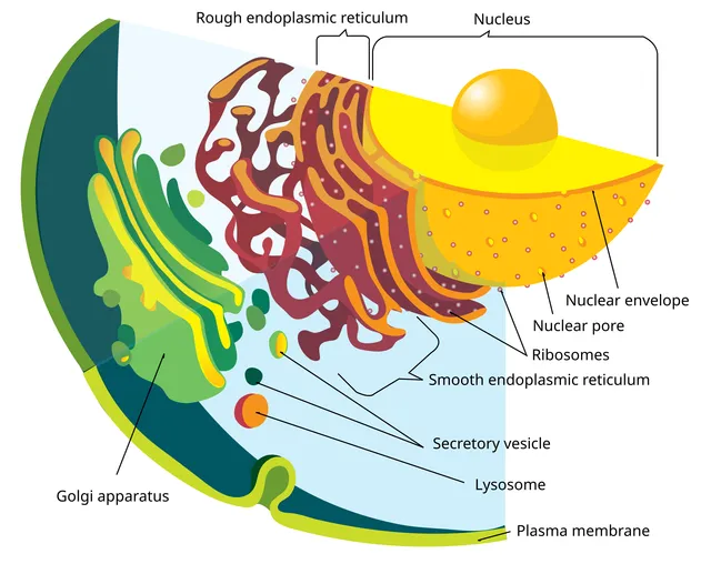
Every protein starts being synthesized on a free ribosome. A signal peptide at the head of the chain diverts the complex to the rough endoplasmic reticulum: the protein continues its synthesis while entering the lumen.
Without a signal peptide, translation finishes in the cytosol, and the protein stays there or goes to the nucleus, mitochondrion or peroxisome according to other targeting sequences.

From the reticulum to the final destination
The reticulum handles folding and the first glycosylations. Correctly folded proteins leave in vesicles for the Golgi, which continues the modifications and above all sorts them.
Three destinations leave from the trans-Golgi network: the plasma membrane through constitutive secretion, regulated secretory granules, and the lysosome.

The mitochondrion
The mitochondrion has two membranes. The outer one is permeable to small molecules; the inner one, highly folded into cristae, is impermeable and carries the respiratory chain.
It has its own circular DNA and its own ribosomes — remnants of its bacterial origin. This DNA is passed on exclusively by the mother.
The secretory pathway, step by step
A protein destined to leave the cell starts its translation on a free ribosome, like all the others. The first amino acids it produces form a signal peptide, recognized on the fly by a signal recognition particle that pauses translation and brings the ribosome to the endoplasmic reticulum.
Translation then resumes through a pore, and the growing chain drops straight into the lumen of the reticulum. There it is folded, often glycosylated, and checked. A misfolded protein does not leave: it is retrotranslocated to the cytosol and destroyed by the proteasome.
Trafficking continues by vesicle budding toward the Golgi apparatus, which the protein crosses from the cis face to the trans face, undergoing different modifications in each cisterna. The trans-Golgi network finally does the sorting: plasma membrane, lysosome, or a secretory granule waiting for a signal.
The bacterial origin of the mitochondrion
The mitochondrion has circular DNA, ribosomes resembling those of bacteria, a double membrane, and it divides by fission rather than being built from scratch. These four features led to the endosymbiotic hypothesis: an ancestral cell, related to the archaea, once engulfed a bacterium without digesting it.
This origin has practical consequences. Antibiotics that target the bacterial ribosome — aminoglycosides, chloramphenicol, linezolid — also reach the mitochondrial ribosome, which accounts for part of their toxicity. And mitochondrial DNA, lacking histones and poorly repaired, mutates about ten times faster than nuclear DNA.
It is passed on exclusively through the mother: the few mitochondria brought by the sperm are tagged and destroyed after fertilization. A disease caused by a mitochondrial DNA mutation is therefore passed on by the mother to all her children, with a severity that varies with the proportion of mutated mitochondria — heteroplasmy; an affected man does not pass it on.
The nucleus
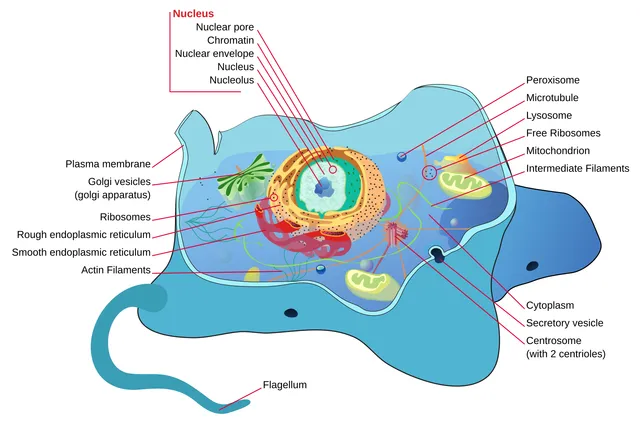
The nucleus is bounded by an envelope made of two membranes, the outer of which is continuous with the endoplasmic reticulum. Beneath the envelope, a network of intermediate filaments, the lamina, gives it shape and anchors the chromatin. The envelope is pierced by nuclear pores, large protein complexes that let small molecules pass freely and control the passage of large ones: proteins bound for the nucleus carry a nuclear localisation signal, and messenger RNAs leave only once processed.
DNA is associated there with proteins, the histones, to form chromatin. Euchromatin, loosely packed, contains active genes; heterochromatin, dense, contains silent regions. The degree of packing is therefore part of gene regulation.
The nucleolus, a dense zone without a membrane, is the ribosome factory: ribosomal RNAs are transcribed there and assembled with proteins imported from the cytosol. Its size reflects the cell's protein-synthesis activity.
Smooth and rough endoplasmic reticulum
The rough endoplasmic reticulum, studded with ribosomes, makes proteins destined for secretion, insertion into a membrane or delivery to lysosomes. In its lumen, the newly made chains receive their first sugars and fold with the help of chaperone proteins. Those that remain misfolded are sent back to the cytosol and destroyed by the proteasome.
The smooth endoplasmic reticulum, without ribosomes, synthesises membrane lipids and steroid hormones. In the hepatocyte, it contains the cytochromes that detoxify many drugs; in the muscle fibre, it is called the sarcoplasmic reticulum and stores the calcium that triggers contraction.
Free ribosomes in the cytosol make the proteins that stay in the cytosol and those destined for the nucleus, mitochondria and peroxisomes, which are imported once complete.
Golgi, lysosomes and storage diseases
The Golgi apparatus is a stack of cisternae that receives proteins from the reticulum on its cis face and dispatches them from its trans face. On the way, it completes their glycosylation and sorts them by destination: plasma membrane, secretion or lysosomes.
Enzymes destined for lysosomes receive a tag in the Golgi, mannose-6-phosphate, recognised by a receptor that directs them to the lysosomal compartment. Lysosomes, kept at a pH of about five by a proton pump, contain hydrolases active in acid conditions, which digest material coming from outside by endocytosis or from the cell itself by autophagy.
When a lysosomal enzyme is missing, its substrate accumulates: these are the lysosomal storage diseases. In Gaucher disease, a lipid accumulates in the macrophages of the liver, spleen and marrow; in Tay-Sachs disease, lipids accumulate in neurons, with early neurological degeneration. If it is the mannose-6-phosphate tag that is missing, all lysosomal enzymes are secreted instead of being delivered, and lysosomes fill with debris.
Diseased mitochondria and peroxisomes
The mitochondrion has its own circular DNA, which encodes a few proteins of the respiratory chain. It is transmitted only by the mother, since the sperm's mitochondria are eliminated after fertilisation. A cell contains many copies of this DNA, normal and mutated in variable proportions: this is heteroplasmy, which explains the great variability of mitochondrial diseases. They mainly affect the most energy-hungry tissues: muscle, brain, retina and heart.
The peroxisome, a small organelle bounded by a single membrane, notably oxidises very-long-chain fatty acids, too long for the mitochondrion, and produces hydrogen peroxide, which it destroys immediately with its catalase. In X-linked adrenoleukodystrophy, these fatty acids cannot enter the peroxisome: they accumulate in the white matter of the brain and in the adrenal gland.
58 answered questions go with this chapter in the practice section.
Sources and further reading
Reference textbooks
- Bruce Alberts et coll., Biologie moléculaire de la cellule.
Open access
- 4.4 The Endomembrane System and Proteins — OpenStax, Biology 2e.
- 3.2 The Cytoplasm and Cellular Organelles — OpenStax, Anatomy and Physiology 2e.
Last revised : . Text licensed under CC BY-SA 4.0.