Cytoskeleton and junctions
Microfilaments, microtubules, intermediate filaments and adhesion.
Three networks, three jobs
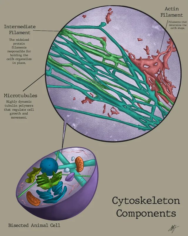
Actin microfilaments, the thinnest at seven nanometers, form a cortex beneath the membrane and govern the cell's shape, its movements and its division ring. They are polarized and constantly remodeled.
Microtubules, the thickest at twenty-five nanometers, are tracks. Kinesin walks toward the plus end, dynein toward the minus end; mitochondria, vesicles and chromosomes move along them. The mitotic spindle is nothing more than a reorganized microtubule network.
Intermediate filaments, in between at ten nanometers, serve neither transport nor movement. They are not polarized, bind neither ATP nor GTP to assemble, and resist traction. Their composition varies with the tissue — keratins in epithelia, vimentin in connective tissue, neurofilaments in neurons — which makes them a valuable marker for identifying the origin of a tumor.

Cell junctions
An epithelium is not a pile of independent cells: it is stitched together. Tight junctions encircle the apical pole and seal the intercellular space, forcing solutes to pass through the cells rather than slip between them. They also maintain polarity, by stopping apical proteins from drifting to the basal side.
Adherens junctions and desmosomes provide mechanical cohesion, the former anchored to actin, the latter to intermediate filaments. Hemidesmosomes do the same job toward the basal lamina.
Gap junctions, finally, open a direct channel between two cytoplasms. Ions and small molecules flow through them, which couples cells electrically — this is what lets the myocardium contract as a single unit.
Cilia, flagella and the centrosome
The centrosome, near the nucleus, is the main organising centre for microtubules. It contains two centrioles, each made of nine microtubule triplets, and duplicates once per cycle: its two copies form the poles of the mitotic spindle.
Motile cilia and the sperm flagellum are built on an axoneme of nine peripheral microtubule doublets arranged around a central pair — the so-called 9 + 2 structure. Dynein arms link neighbouring doublets; by sliding them past one another, they bend the axoneme and produce the beat.
The cilia of the respiratory epithelium beat in coordination and carry mucus up towards the pharynx; those of the uterine tube move the oocyte towards the uterus.
Actin at work
Actin exists in two forms: free monomers in the cytosol and polarised filaments, which grow at one end and shrink at the other. By regulating this turnover, the cell builds and dismantles its filaments within seconds.
At the edge of a moving cell, actin polymerises against the membrane and pushes it forwards, forming flat sheets, lamellipodia, or thin projections, filopodia. Together with myosin, it forms contractile cables that pull the rear of the cell. This is how white cells migrate towards an infection or skin cells towards a wound.
Actin also forms the core of the enterocyte's microvilli, and the contractile ring that pinches the cell in two at the end of division, during cytokinesis. In these roles it works with a non-muscle myosin, close to that of muscle.
Microtubules in action
Microtubules are hollow tubes of tubulin, polarised, whose minus end is anchored in the centrosome while the plus end explores the cytoplasm. They constantly alternate between growth and sudden shrinkage: this is dynamic instability, which lets them search for and capture their targets, such as chromosomes during mitosis.
They serve as rails for molecular motors. Kinesin usually moves towards the plus end, from the cell body to the periphery; dynein moves towards the minus end, from the periphery to the centre. In the axon, sometimes a metre long, this transport carries proteins and organelles in both directions.
Several drugs act on microtubules. Colchicine prevents their assembly and slows the migration of neutrophils, which makes it a treatment for gout attacks. Vinca alkaloids depolymerise them and taxanes freeze them: in both cases the spindle stops working, and cancer cells, which divide a lot, are the first to be affected.
Intermediate filaments and tissue identity
Intermediate filaments are strong, non-polarised ropes that absorb tension. Their nature changes with the tissue: keratins in epithelia, vimentin in cells of mesenchymal origin, desmin in muscle, neurofilaments in neurons, glial fibrillary acidic protein in astrocytes, and lamins in the nucleus of every cell.
This specificity makes them valuable markers. Faced with a metastasis of unknown origin, the pathologist uses immunohistochemistry to look for the intermediate filaments expressed: keratins point to a carcinoma, of epithelial origin, vimentin to a sarcoma, and the glial protein to a brain tumour.
Their failure weakens tissues under stress. A mutation in the keratins of the basal layer of the epidermis makes the skin so fragile that simple rubbing lifts it into blisters: this is a form of epidermolysis bullosa.
When junctions come under attack
In pemphigus, the immune system produces antibodies against desmogleins, the cadherins of epidermal desmosomes. Keratinocytes come apart from one another: fragile blisters form within the epidermis, which rupture leaving erosions, including in the mouth.
In bullous pemphigoid, more common in older people, the antibodies target hemidesmosomes, which attach the epidermis to the basement membrane. The whole epidermis lifts off the dermis: the blisters, beneath the epidermis, are tense and firm. The depth of the blister, visible under the microscope, distinguishes the two diseases.
Junctions also play a part in cancer. Loss of the cadherin that binds epithelial cells together makes it easier for them to detach from the tumour, migrate and form metastases. And gap junctions, made of connexins, are essential to the inner ear: a mutation in one of them is the most common genetic cause of congenital deafness.
58 answered questions go with this chapter in the practice section.
Sources and further reading
Reference textbooks
- Bruce Alberts et coll., Biologie moléculaire de la cellule.
Open access
- 4.5 The Cytoskeleton — OpenStax, Biology 2e.
- 4.6 Connections between Cells and Cellular Activities — OpenStax, Biology 2e.
Last revised : . Text licensed under CC BY-SA 4.0.