From DNA to protein
Transcription, processing, translation and the genetic code.
The genetic code
The genetic code assigns an amino acid to each triplet of bases. It is redundant — several codons for the same amino acid — but unambiguous: a given codon never designates two. It is read without overlap, from an initiating AUG, up to one of the three stop codons.
Degeneracy mainly concerns the third base, which limits the impact of point mutations at that position.
From gene to protein
In eukaryotes, transcription and translation are separated in space and time. RNA polymerase II transcribes the gene in the nucleus into a pre-messenger RNA, which must then be processed before it can leave.
Three operations transform it. A cap is added to the 5' end, which protects against degradation and serves as the docking point for the ribosome. A poly-adenosine tail is added at the 3' end, which stabilizes it. And above all, the introns are removed by the spliceosome, the exons being joined end to end.
This last step is not mere cleanup. By keeping or leaving out certain exons, a single cell produces several different messengers from a single gene, and therefore several proteins. Alternative splicing largely explains the gap between the roughly twenty thousand human protein-coding genes and the far greater diversity of proteins produced.

Reading the code, and what happens when it goes wrong
A single-base substitution can therefore have very unequal consequences. If the new codon designates the same amino acid, the mutation is silent. If it designates another, it is missense, and its severity depends on how chemically similar the two are. If it creates a stop codon, it is nonsense and the protein is truncated.
Inserting or deleting a number of bases that is not a multiple of three is of a different nature: it shifts the reading frame, and every codon downstream changes. The result is almost always an unrecognizable protein, often cut short prematurely.
Transcription in detail
In eukaryotes, three RNA polymerases share the work. RNA polymerase I makes most ribosomal RNAs, in the nucleolus; RNA polymerase II transcribes protein-coding genes into pre-messenger RNAs, as well as many small regulatory RNAs; RNA polymerase III makes transfer RNAs and the smallest ribosomal RNA.
RNA polymerase II does not recognise the promoter on its own. General transcription factors first bind sequences near the start site, such as the TATA box, then recruit the polymerase. More distant sequences, enhancers, sometimes tens of thousands of base pairs away, bind activator proteins that greatly increase transcription by looping the DNA to reach the promoter.
The polymerase reads the template strand in the 3' to 5' direction and synthesises RNA in the 5' to 3' direction. The RNA therefore has the sequence of the non-template strand, called the coding strand, except that thymine is replaced by uracil. Unlike DNA polymerase, RNA polymerase needs no primer.
Splicing and its errors
Most human genes are split: their coding parts, the exons, are separated by introns, often much longer. Splicing removes the introns from the pre-messenger RNA and joins the exons together. It is carried out by the spliceosome, a large complex of proteins and small nuclear RNAs, which recognises short signals at each end of the intron: almost always GU at the start and AG at the end.
A mutation that destroys one of these signals, or creates a new one, disrupts splicing: an exon may be skipped, or a piece of intron retained. Some beta-thalassaemias are an example: a single base changed in an intron of the beta-globin gene creates a false splice site and prevents production of a normal chain. A substantial share of disease-causing mutations are thought to act through this mechanism.
Alternative splicing, where one pre-messenger is spliced in several ways, affects the great majority of multi-exon human genes. It allows different proteins to be produced depending on tissue or timing, for example membrane-bound and secreted forms of the same antibody.
The ribosome at work
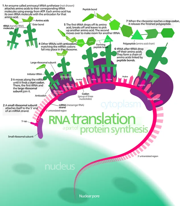
The eukaryotic ribosome, called 80S, combines a small 40S subunit and a large 60S subunit, made of ribosomal RNAs and proteins. It is the RNA of the large subunit that catalyses peptide bond formation: the ribosome is a ribozyme. It has three sites for transfer RNAs: the A site receives the tRNA carrying the next amino acid, the P site holds the tRNA bearing the growing chain, the E site lets the empty tRNA leave.
Initiation begins when the small subunit, bound near the cap, slides along the messenger to the first AUG codon in a favourable context. An initiator tRNA carrying methionine pairs with it, then the large subunit joins. At each elongation step, a new tRNA enters the A site, the chain is transferred onto its amino acid, and the ribosome moves on by one codon.
Termination occurs when a stop codon, UAA, UAG or UGA, reaches the A site. No tRNA recognises it: a release factor binds and causes the chain to detach. A single messenger is read simultaneously by several ribosomes, which form a polysome.
The bacterial ribosome, called 70S, made of 30S and 50S subunits, differs enough from the human ribosome for antibiotics to block it selectively: aminoglycosides and tetracyclines target the small subunit, macrolides the large one.
After translation
A protein's destination is often written in its sequence. Proteins destined for secretion, the plasma membrane or lysosomes begin with a hydrophobic signal peptide: as soon as it emerges from the ribosome, it is recognised and the ribosome is brought to the endoplasmic reticulum, where the chain is fed in as it is made. Proteins without a signal stay in the cytosol or reach the nucleus and mitochondria through other tags.
The chain must then fold, often with the help of chaperone proteins. It undergoes modifications that change its activity or destination: glycosylation in the reticulum and Golgi apparatus, formation of disulfide bridges, phosphorylation that switches an enzyme on or off, cleavage of a precursor, such as proinsulin into insulin.
Misfolded or no longer needed proteins are tagged with chains of ubiquitin and destroyed by the proteasome. Some neurodegenerative diseases involve aggregates of misfolded proteins that this system failed to clear.
Chromatin and epigenetic memory
Nuclear DNA is wound around histone octamers, forming nucleosomes. Euchromatin, loosely packed, contains active genes; heterochromatin, densely packed, is silent. Marks placed on DNA and histones, without changing the sequence, determine which genes are accessible: this is epigenetics.
Methylation of cytosines followed by a guanine, in the CG-rich islands of promoters, generally switches the gene off. It is copied at each replication by a maintenance enzyme, which passes the gene's state on to daughter cells: a liver cell stays a liver cell. Histone acetylation, which loosens chromatin, goes along instead with active transcription.
X inactivation in females is the most visible illustration. Early in the embryo, each cell randomly silences one of its two X chromosomes, which condenses into a Barr body; the choice is then passed on to all its descendants. A woman is therefore a mosaic of territories expressing either the paternal or the maternal X, which explains why carriers of an X-linked disease sometimes show signs, depending on the proportion of cells in which the mutated X is active.
58 answered questions go with this chapter in the practice section.
Sources and further reading
Reference textbooks
- Bruce Alberts et coll., Biologie moléculaire de la cellule.
- Robert L. Nussbaum, Roderick R. McInnes, Huntington F. Willard, Thompson & Thompson. Génétique médicale, Elsevier Masson.
Open access
- 15.1 The Genetic Code — OpenStax, Biology 2e.
- 15.5 Ribosomes and Protein Synthesis — OpenStax, Biology 2e.
Last revised : . Text licensed under CC BY-SA 4.0.