DNA replication and repair
Semiconservation, the fork, fidelity and repair systems.
Semiconservative replication
Each strand serves as a template for the synthesis of its complement: the two daughter molecules each contain an old strand and a new one. This is semiconservation, demonstrated by the Meselson-Stahl experiment.
DNA polymerase only synthesizes in the 5' to 3' direction. One strand is therefore copied continuously, the other as successive fragments that are joined afterward — hence the asymmetry of the fork.

Fidelity
The raw error rate of base pairing would be very high. It is brought down to an extremely low level by two mechanisms: the proofreading activity of the polymerase itself, then the mismatch repair that follows synthesis.
Other systems repair chemical lesions: base excision, nucleotide excision, double-strand break repair. Their inherited failure shows up as cancer predisposition syndromes.
An asymmetric fork, and why
DNA polymerase can only do one thing: extend a free 3' end. It can therefore only synthesize in the 5' to 3' direction, and it needs a primer to start, supplied by a primase in the form of RNA.
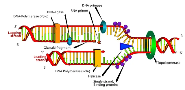
But the two strands of the double helix are antiparallel. At the replication fork, one runs in the right direction and can be copied continuously: this is the leading strand. The other runs the wrong way and can only be copied in short successive stretches, each with its own primer: these are the Okazaki fragments of the lagging strand.
The RNA primers are then removed, the gaps filled, and the fragments joined by a ligase. All the asymmetry of the fork comes from a single enzymatic constraint.
Fidelity and repair
DNA polymerase makes a mistake about once in a hundred thousand, but it proofreads: its exonuclease activity immediately removes a mismatched nucleotide. The error rate then drops to one in ten million.
A second system, mismatch repair, follows behind and corrects what slipped through. Its inherited deficiency causes Lynch syndrome, which predisposes to colorectal cancer.
Other systems deal with lesions, which are distinct from copying errors. Nucleotide excision removes the thymine dimers induced by ultraviolet light; its deficiency causes xeroderma pigmentosum, with skin cancers from childhood. Base excision deals with point chemical alterations. Homologous recombination repairs double-strand breaks, and the BRCA genes are part of it.
The team at the fork
At the replication fork, a helicase separates the two strands by breaking the hydrogen bonds between bases, using ATP. Single-stranded DNA-binding proteins immediately attach to prevent the strands from re-pairing. Ahead of the fork, topoisomerases briefly cut the DNA to relieve the twisting that builds up.
No DNA polymerase can start on its own: it needs a free 3' end. Primase, which is an RNA polymerase, therefore lays down a short RNA primer. The replicative DNA polymerases then extend the chain, always in the 5' to 3' direction, held on the DNA by a sliding clamp, PCNA, which lets them copy long stretches without falling off.
On the lagging strand, Okazaki fragments are made one after another. Their RNA primers are removed and replaced with DNA, then DNA ligase seals the fragments together.
Topoisomerases are drug targets. Fluoroquinolones, which are antibiotics, block the bacterial type II topoisomerase, DNA gyrase. Several anticancer drugs block human topoisomerases and leave breaks in the DNA of dividing cells.
Replicating a human genome
The human genome has about three billion base pairs per haploid set. A fork moves at about fifty nucleotides per second in humans: with a single origin, replication would take weeks. It actually starts at tens of thousands of origins, which allows it to be completed in an S phase of a few hours.
Each origin must be used only once per cycle. In G1, loading proteins "license" it by installing the inactive helicase; at the start of S phase, cell-cycle kinases activate the helicase and at the same time prevent any new loading. DNA is thus copied once, no more and no less.
At the ends of chromosomes, the last fragment of the lagging strand cannot be completed after its primer is removed: DNA shortens with each division. Telomeres, repeats of a short TTAGGG sequence, provide a margin. Telomerase, which carries its own RNA template, lengthens them in stem and germ cells; mutations that weaken it cause diseases in which bone marrow, skin and lungs wear out prematurely.
Where damage comes from
DNA is constantly damaged, even without external attack. Each cell loses thousands of purine bases every day through spontaneous breakage of their bond to the sugar, depurination. Cytosine turns spontaneously into uracil through deamination; if this change is not repaired, a G-C pair becomes A-T at the next replication. Reactive oxygen species, produced by mitochondrial respiration, oxidise bases, notably guanine.
External agents add their share. Ultraviolet light links two neighbouring pyrimidines on the same strand, particularly two thymines, forming dimers that distort the double helix. Ionising radiation breaks both strands at once. Alkylating agents, including some anticancer drugs, attach groups to bases; compounds in tobacco smoke form bulky adducts.
A lesion is not yet a mutation: it is a chemical abnormality that the cell can repair. It becomes a mutation when replication copies it incorrectly before repair, and the error is then passed on to both daughter cells.
The repair systems
Each type of lesion has its system. Base excision removes a single damaged base, uracil or oxidised guanine, using glycosylases that recognise it; the gap is filled and sealed. Nucleotide excision removes a segment of about thirty nucleotides around a lesion that distorts the helix, such as a thymine dimer. Its inherited defect causes xeroderma pigmentosum: extreme sensitivity to sunlight and multiple skin cancers from childhood.
Mismatch repair corrects errors left by the polymerase, by recognising the new strand. An inherited defect in its genes causes Lynch syndrome, which predisposes to cancers of the colon and endometrium; the tumours show microsatellite instability, short repeats whose length varies for lack of correction.
Double-strand breaks, the most dangerous, have two pathways. Homologous recombination uses the sister chromatid as a template: it is accurate but is possible only after replication, in S and G2. It depends notably on BRCA1 and BRCA2, whose inherited mutations predispose to breast and ovarian cancer. Non-homologous end joining sticks the ends directly back together, at any point in the cycle, at the cost of small losses.
Tumours lacking BRCA survive thanks to other repair pathways. PARP inhibitors block one of them: the tumour cells, left with no solution, die, whereas normal cells, which keep BRCA, tolerate the treatment. This is the principle of synthetic lethality.
Copying DNA in the laboratory
PCR copies a chosen DNA segment by repeating three steps. At about 95 °C, the strands separate; at around 55 to 65 °C, two short primers anneal on either side of the target sequence; at about 72 °C, a heat-resistant polymerase, from a hot-spring bacterium, extends the primers. Each cycle doubles the number of copies: thirty cycles produce about a billion.
Sanger sequencing reads a sequence by adding to the replication mixture modified nucleotides, lacking a 3'-OH group, which stop synthesis. Fragments of every length are obtained, each ending in a base labelled with a colour; sorting them by size gives the sequence.
High-throughput sequencing techniques read millions of fragments in parallel. They allow the exome, all the exons, or the whole genome of a patient to be analysed at once, and have transformed the diagnosis of rare genetic diseases.
58 answered questions go with this chapter in the practice section.
Sources and further reading
Reference textbooks
- Bruce Alberts et coll., Biologie moléculaire de la cellule.
Open access
- 14.5 DNA Replication in Eukaryotes — OpenStax, Biology 2e.
- 14.6 DNA Repair — OpenStax, Biology 2e.
Last revised : . Text licensed under CC BY-SA 4.0.