Meiosis and genetic shuffling
Two divisions, crossing over, shuffling and anomalies.
Two divisions, a single replication
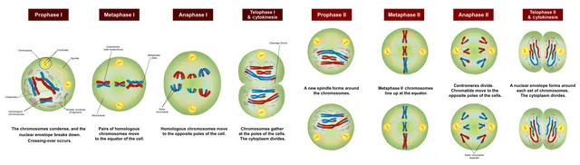
Meiosis runs two divisions after a single round of replication: one diploid cell gives four haploid cells. This is chromosome reduction, essential so that fertilization restores the original number.
The first division separates homologous chromosomes — it is the reductional one; the second separates sister chromatids, like a mitosis.

Two kinds of shuffling, and the count of combinations
Meiosis produces diversity through two distinct mechanisms, which must not be confused.
Intrachromosomal shuffling is crossing over: in prophase I, non-sister chromatids of two homologs exchange segments. A single chromosome then carries a mixture of paternal and maternal alleles that neither origin contained.
Interchromosomal shuffling comes from the orientation of each pair on the equatorial plate in metaphase I. Each of the twenty-three pairs orients independently of the others, which gives two to the power of twenty-three combinations, more than eight million — before any crossing over at all.
When separation fails
A nondisjunction is the failure of two chromosomes that should have separated to do so. In anaphase I, it is the homologs that stay together; in anaphase II, the sister chromatids. In both cases, one gamete receives an extra chromosome and the other one fewer.
Once fertilized, the first gives a trisomy, the second a monosomy. Most are lethal early; a few are viable, trisomy 21 foremost among them.
The risk rises sharply with maternal age, and the mechanism is illuminating: the oocyte is arrested in prophase I from fetal life and only completes its first division at ovulation. In a forty-year-old woman, that wait has lasted forty years, and the proteins holding the chromosomes paired have degraded.
Prophase I, stage by stage
Prophase of the first division is long and divided into five stages. At leptotene, the already replicated chromosomes begin to condense into thin threads. At zygotene, each chromosome pairs with its homologue along its whole length, thanks to a zipper-like protein structure, the synaptonemal complex: each pair forms a bivalent, made of four chromatids.
At pachytene, chromosomes are fully paired and this is when crossing-over occurs: exchanges of segments between non-sister chromatids. At diplotene, the synaptonemal complex comes apart and the homologues move away from each other, except where a crossover has occurred, which remain visible as chiasmata. It is at diplotene that the oocyte arrests for years, in a state called dictyotene.
At diakinesis, chromosomes reach maximum condensation and the nuclear envelope disappears. Chiasmata are not just a trace: they hold the homologues together until anaphase I. Each bivalent must have at least one for the two homologues to separate correctly; a bivalent with no chiasma is at risk of non-disjunction.
Mitosis and meiosis side by side
The two divisions share the mechanics of the spindle, but their logic differs. Mitosis conserves: one diploid cell gives two identical diploid cells. Meiosis reduces and shuffles: one diploid cell gives four haploid cells, all different.
The decisive difference lies in anaphase I. In mitosis, sister chromatids separate. In anaphase I of meiosis, it is the homologous chromosomes that separate, each keeping its two sister chromatids joined: cohesins are cut only along the arms, not at the centromere. The second division then resembles a mitosis, but on haploid cells and without prior replication.
- Mitosis: one division; meiosis: two divisions for a single replication.
- Mitosis: no pairing of homologues; meiosis: pairing and crossing-over in prophase I.
- Mitosis: 2 cells at 2n; meiosis: 4 cells at n.
- Mitosis: identical daughter cells; meiosis: genetically different daughter cells.
- Mitosis: somatic cells; meiosis: germ line.
Aneuploidies
An aneuploidy is an abnormal number of chromosomes, too many or too few. Most autosomal monosomies and trisomies are lethal very early and account for a large share of first-trimester miscarriages. Three autosomal trisomies are compatible with birth: trisomy 21, by far the most common, and trisomies 18 and 13, which are much more severe.
In about 95% of cases, trisomy 21 is free: three separate chromosomes 21, resulting from a non-disjunction, most often during maternal meiosis and mainly in the first division. More rarely, the extra chromosome 21 is attached to another by a Robertsonian translocation; the parents' karyotype must then be done, as one of them may carry the translocation in balanced form and the recurrence risk is higher. Finally, a non-disjunction occurring after fertilisation gives a mosaic, with normal and trisomic cells, and often a milder picture.
For the sex chromosomes, deviations are better tolerated, thanks to inactivation of the extra X and the few genes carried by Y. Turner syndrome, 45,X, the only viable monosomy, combines short stature and non-functioning ovaries; the missing chromosome is most often of paternal origin. Klinefelter syndrome, 47,XXY, combines tall stature, small testes and infertility.
Structural abnormalities
A reciprocal translocation exchanges segments between two non-homologous chromosomes. If no material is lost, it is called balanced and the carrier is healthy. But at meiosis, the rearranged chromosomes and their normal homologues must pair in a cross shape, and some gametes receive an unbalanced set: the carrier has an increased risk of miscarriage or of a child with an abnormality.
A Robertsonian translocation fuses two acrocentric chromosomes, whose centromere lies near one end — chromosomes 13, 14, 15, 21 and 22 — by their long arms, the short arms being lost without consequence. The balanced carrier has only 45 chromosomes but all their useful genetic material.
A deletion removes a segment: that of the short arm of chromosome 5 causes cri du chat syndrome. Microdeletions, too small to be seen on a standard karyotype, such as the 22q11 deletion, are sought by fluorescence in situ hybridisation, where a labelled probe reveals the presence or absence of a precise region, or by chromosomal microarray analysis, which detects gains and losses across the whole genome.
Counting the combinations
Interchromosomal shuffling relies on the random orientation of each pair of homologues at metaphase I. With 23 pairs, an individual can produce 2 to the power 23 combinations of chromosomes, about 8.4 million different gametes, before even counting crossovers.
For a couple, the number of possible different zygotes is the product of the two: about 8.4 million squared, some 70 trillion. Crossovers, several dozen per meiosis, multiply this number further to the point that, in practice, two gametes from the same individual are never identical. Only monozygotic twins share the same genome.
58 answered questions go with this chapter in the practice section.
Sources and further reading
Reference textbooks
- Robert L. Nussbaum, Roderick R. McInnes, Huntington F. Willard, Thompson & Thompson. Génétique médicale, Elsevier Masson.
- Bruce Alberts et coll., Biologie moléculaire de la cellule.
Open access
- 11.1 The Process of Meiosis — OpenStax, Biology 2e.
Last revised : . Text licensed under CC BY-SA 4.0.