Modes of inheritance
Dominance, recessiveness, X-linked and mitochondrial inheritance.
Autosomal dominant or recessive
Autosomal dominant inheritance shows up in every generation, affects both sexes, and an affected person has a one-in-two chance of passing on the allele to each child.
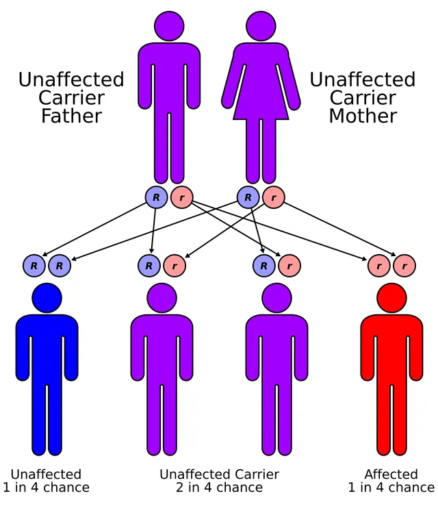
Autosomal recessive inheritance skips generations: the parents are healthy carriers, and the risk for each child of a carrier couple is one in four. Consanguinity markedly increases its frequency.

Special modes of inheritance
In X-linked recessive inheritance, boys are affected and girls are most often healthy carriers: a boy has only one X, and therefore no compensating allele. An affected father never passes it to his sons, to whom he only gives his Y.
Mitochondrial inheritance is maternal: a mother passes her mitochondrial DNA to all her offspring — with variable expression depending on the degree of heteroplasmy — while an affected father passes it to none of his children.
Reading a pedigree
Faced with a pedigree, a few questions are enough to point the way. Does the disease affect both sexes equally? Does it appear in every generation, or does it skip? Is there father-to-son transmission?
Continuous vertical transmission, affecting both sexes, with a one-in-two risk for each child of an affected person, suggests autosomal dominance. Isolated cases born to unaffected parents, often in a context of consanguinity, suggest autosomal recessiveness.
Almost exclusive involvement of boys, passed on through unaffected mothers, suggests X-linked recessiveness. One argument is decisive: father-to-son transmission is impossible, since the father only passes his Y chromosome to his sons. Conversely, all the daughters of an affected man receive his X and are therefore carriers.
Calculating a risk
The calculation relies on the Punnett square: write the possible gametes of each parent and combine them. For two parents heterozygous for an autosomal recessive disease, written Aa, children are AA for a quarter, Aa for half, aa for a quarter: the risk of an affected child is one in four at each pregnancy, regardless of earlier pregnancies.
One subtlety comes up often. An unaffected brother or sister of an affected child cannot be aa: three squares remain, two of them heterozygous. Their probability of being a carrier is therefore two thirds, not one half.
To estimate this person's risk of in turn having an affected child, three probabilities must be multiplied: that they are a carrier, two thirds; that their partner from the general population is one, for example one in thirty for cystic fibrosis in France; that the child receives both mutated alleles, one quarter. The product, 2/3 × 1/30 × 1/4, equals one in one hundred and eighty.
Diseases that serve as models
Cystic fibrosis, autosomal recessive, is the most common serious genetic disease in people of European descent. The CFTR gene codes for a chloride channel; its defect thickens secretions in the airways, pancreas and gut. Sickle cell disease, also recessive, is the most common genetic disease in France, especially in people originating from Africa, the Caribbean and the Mediterranean basin; it is screened for at birth.
Huntington's disease, autosomal dominant, is due to an abnormally long repeat of the CAG triplet. Signs — involuntary movements, psychiatric disturbance, cognitive decline — usually begin around forty, often after the person has had children. The repeat tends to lengthen from one generation to the next, especially when passed on by the father, and the disease then begins earlier: this is anticipation.
Neurofibromatosis type 1, dominant, illustrates variable expressivity: café-au-lait spots, small skin tumours, sometimes serious complications, with huge differences between members of the same family. About half of cases are due to a new mutation, absent in both parents. Haemophilia A and Duchenne muscular dystrophy are examples of X-linked recessive inheritance.
What blurs the pedigrees
Several phenomena make a family tree look unlike the expected pattern. Incomplete penetrance means that a carrier of a dominant mutation remains unaffected: the disease seems to skip a generation. Variable expressivity means that carriers are affected to very different degrees. A new mutation makes a dominant disease appear in a child of healthy parents.
Germline mosaicism is even more misleading: a healthy parent carries the mutation in only some of their germ cells. They can have several children affected by a dominant disease while their own blood shows nothing. This is why the recurrence risk after a first child with an apparently new mutation is never quite zero.
Genetic heterogeneity complicates diagnosis. Mutations in different genes can cause the same disease, which is locus heterogeneity, as in many forms of deafness; different mutations in the same gene can cause different diseases, which is allelic heterogeneity. Finally, dominance is not always clear-cut: for blood groups A and B, both alleles are expressed together in an AB person, which is codominance.
When the parent of origin matters
For a few hundred genes, only the copy from one parent is active, the other being silenced by methylation in the germ line: this is genomic imprinting. The same abnormality then causes different diseases depending on the parent of origin.
The 15q11-q13 region is the classic example. If the paternal region is missing or not working, the child has Prader-Willi syndrome: floppiness at birth, then insatiable appetite and obesity. If the maternal region is missing, the child has Angelman syndrome: severe intellectual disability, absent speech, frequent laughter, epilepsy.
These syndromes can also result from uniparental disomy: the child does have two chromosomes 15, but both come from the same parent. Two maternal chromosomes 15, with no active paternal copy, thus cause Prader-Willi syndrome without any deletion.
Genetic counselling
Genetic counselling informs a person or couple about an inherited risk and the possible choices. It relies on a careful family tree, a precise diagnosis in the family and, where possible, identification of the mutation.
Presymptomatic testing looks for the mutation in a person who is still healthy, for a disease that will appear later, such as Huntington's disease. Because some of these diseases have no preventive treatment, it follows a supervised protocol: repeated consultations, psychological support, a period for reflection, and the person can at any time decide not to learn the result. A minor is tested only if there is a direct medical benefit to them.
When a serious genetic abnormality is found in a person, and prevention or care is available for their relatives, French law requires them to inform those relatives, either themselves or through the doctor, who passes on the information without revealing the person's name or result.
58 answered questions go with this chapter in the practice section.
Sources and further reading
Reference textbooks
- Robert L. Nussbaum, Roderick R. McInnes, Huntington F. Willard, Thompson & Thompson. Génétique médicale, Elsevier Masson.
Open access
- 12.3 Laws of Inheritance — OpenStax, Biology 2e.
- 13.1 Chromosomal Theory and Genetic Linkage — OpenStax, Biology 2e.
Last revised : . Text licensed under CC BY-SA 4.0.