Connective tissues
Extracellular matrix, fibers, cells and varieties.
Very different varieties
The same organizational plan gives tissues with opposite properties depending on the nature and arrangement of the matrix: loose connective tissue fills and nourishes, tendon resists traction, cartilage resists compression, bone bears load, adipose tissue stores, blood transports.
A matrix, a few cells
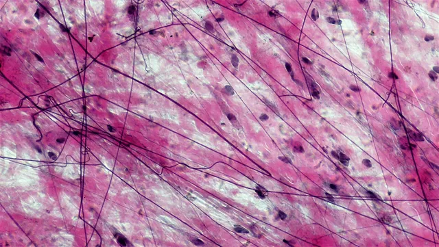
Unlike an epithelium, a connective tissue is made mostly of extracellular matrix, with cells scattered within it. This matrix comprises fibers and a ground substance.
Collagen fibers, resistant to traction, dominate by mass — collagen is the most abundant protein in the body. Elastic fibers, made of elastin, make deformation reversible: they abound in the arterial wall and the lung. Reticular fibers form a fine supporting meshwork in lymphoid organs and the liver.
The ground substance is a highly hydrated gel of proteoglycans and hyaluronic acid. It cushions, slows the spread of bacteria and serves as a water reservoir. Some bacteria secrete a hyaluronidase precisely to thin it out and advance faster.

Bone, a connective tissue that renews itself
Bone is a connective tissue whose matrix is mineralized by hydroxyapatite crystals laid down on a framework of type I collagen. This combination provides both resistance to compression, from the mineral, and resistance to traction, from the framework.
Three cells maintain it. The osteoblast, of mesenchymal origin, secretes the matrix, then lets itself be walled in and becomes an osteocyte. The osteocyte, in its lacuna, senses mechanical loads and signals where reinforcement is needed. The osteoclast, multinucleated and derived from the monocyte lineage, resorbs bone by acidifying the space beneath its ruffled border.
This constant remodeling explains why bone adapts to use — it becomes denser under load and thinner with immobilization — and why it can serve as a calcium reservoir mobilized under the effect of parathyroid hormone.
The cells of connective tissue
Resident cells, which arise and live in the tissue, are distinguished from transient cells coming from the blood. The fibroblast is the main resident cell: it makes fibres and ground substance and renews them. At rest it is sometimes called a fibrocyte; activated by a wound, it multiplies and can turn into a myofibroblast, a contractile cell that pulls the edges of the scar together.
The adipocyte stores triglycerides. The tissue macrophage, derived from a blood monocyte, phagocytoses debris and microbes and presents antigens. The mast cell, packed with granules of histamine and heparin, triggers the immediate inflammatory reaction when IgE bound to its surface recognises an allergen.
Plasma cells, derived from B lymphocytes, produce antibodies; their nucleus with cartwheel chromatin and their abundant reticulum make them recognisable. Granulocytes and lymphocytes leave the vessels in large numbers during inflammation.
From gene to collagen fibre
Each collagen molecule combines three chains wound into a triple helix. One amino acid in three is glycine, the smallest, the only one able to fit at the centre of the helix. In the endoplasmic reticulum, proline and lysine residues are hydroxylated by enzymes that need vitamin C; these hydroxylations stabilise the helix.
The molecule leaves the cell as procollagen, with ends that prevent it from assembling too early. These ends are cut off outside; the molecules then line up into staggered fibrils, which cross-links strengthen, and the fibrils group into fibres.
Each step has its disease. Without vitamin C, collagen is fragile: this is scurvy, with bleeding gums and poorly healing wounds. A mutation of collagen I, often the replacement of a glycine, causes osteogenesis imperfecta and its repeated fractures. Ehlers-Danlos syndromes combine very stretchy skin and overly mobile joints.
Elastic fibres combine a core of elastin and a sheath of fibrillin microfibrils. A fibrillin mutation causes Marfan syndrome: tall stature, long limbs, dislocation of the lens and, above all, dilatation of the aorta that threatens to rupture.
Cartilage, a tissue without vessels
Cartilage is a connective tissue whose matrix, rich in water-binding proteoglycans, resists compression. Its cells, the chondrocytes, sit in cavities called lacunae, often in small groups derived from a single mother cell: isogenous groups.
Cartilage contains neither vessels nor nerves. It is nourished by diffusion from the perichondrium, the connective sheath around it, or from synovial fluid in the case of articular cartilage, which has no perichondrium. It grows in two ways: by division of chondrocytes inside it, interstitial growth, and by laying down new layers from the perichondrium, appositional growth.
There are three varieties. Hyaline cartilage, the most widespread, forms joint surfaces, costal cartilages and the rings of the trachea. Elastic cartilage, rich in elastic fibres, forms the external ear and the epiglottis. Fibrocartilage, rich in collagen I, forms the intervertebral discs, the pubic symphysis and the menisci.
Lacking vessels, articular cartilage repairs its injuries very poorly. In osteoarthritis it gradually thins and cracks, and the underlying bone reacts by becoming denser and forming outgrowths.
Bone: architecture and formation
Compact bone, which forms the cortex of long bones, is built of osteons: cylinders of concentric lamellae around a central canal, the Haversian canal, which contains vessels and nerves. Osteocytes, lodged between the lamellae, communicate through fine canaliculi. Spongy bone, inside the ends of bones and in the vertebrae, is a network of trabeculae between which the bone marrow lies.
Bone forms in two ways. In intramembranous ossification, mesenchymal cells turn directly into osteoblasts: this is how the flat bones of the skull and part of the clavicle arise. In endochondral ossification, a hyaline cartilage model is first formed and then replaced by bone: this applies to most bones, including the long bones.
In children, a disc of cartilage, the growth plate, persists between the epiphysis and the metaphysis. Its chondrocytes line up in columns, enlarge and then die, and their cartilage is replaced by bone: this is how the bone lengthens. At the end of puberty, under the influence of oestrogens in both sexes, the plate ossifies and growth in length stops.
Blood calcium depends on bone. Parathyroid hormone indirectly stimulates resorption by osteoclasts and raises blood calcium; vitamin D promotes intestinal calcium absorption; calcitonin restrains osteoclasts. In osteoporosis, resorption outweighs formation: bone mass and trabecular quality fall, and fractures occur after minimal impacts.
Adipose tissue, an endocrine organ
White adipose tissue is made of adipocytes each containing a single large lipid droplet that pushes the nucleus to the edge. It stores energy, insulates and cushions. It is not inert: it secretes leptin, a hormone that signals abundant reserves to the hypothalamus and reduces appetite. Most people with obesity have high leptin levels but respond poorly to it.
Brown adipose tissue contains adipocytes with many small lipid droplets and very rich in mitochondria. These mitochondria carry an uncoupling protein, thermogenin, which lets protons flow back in without making ATP: the energy is dissipated as heat. Abundant in the newborn, between the shoulder blades and around the great vessels, it protects against cold; small amounts persist in adults.
58 answered questions go with this chapter in the practice section.
Sources and further reading
Reference textbooks
- Alan Stevens, James Lowe, Histologie humaine, Elsevier Masson.
Open access
- 4.3 Connective Tissue Supports and Protects — OpenStax, Anatomy and Physiology 2e.
Last revised : . Text licensed under CC BY-SA 4.0.