Epithelia
Classification, polarity, junctions and renewal.
Basement membrane and nutrition
An epithelium contains no blood vessels. It rests on a basement membrane, a thin layer of matrix that anchors it to the underlying connective tissue, and is nourished by diffusion across it.
Classifying an epithelium in two questions
Every epithelium is described by two features: the number of layers and the shape of the surface cells. With a single layer it is simple; with several, stratified; with a single layer whose nuclei sit at different heights, pseudostratified. The cells are squamous if flattened, cuboidal if as tall as they are wide, columnar if taller than they are wide.
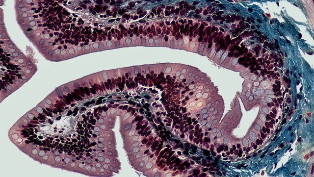
Form follows function. A simple squamous epithelium, thin, favors exchange: it lines the pulmonary alveolus and the vascular endothelium. A stratified squamous epithelium, thick, protects against friction: skin, esophagus, vagina. A simple columnar epithelium, rich in organelles, secretes and absorbs: stomach, intestine.
The urothelium is a case apart. Its umbrella-shaped surface cells flatten when the bladder fills and thicken when it empties, so its appearance changes with the degree of filling — hence its former name of transitional epithelium.
- Pulmonary alveolus, endothelium: simple squamous.
- Renal tubule, gland ducts: simple cuboidal.
- Stomach, intestine, gallbladder: simple columnar.
- Trachea, bronchi: ciliated pseudostratified.
- Skin: keratinized stratified squamous. Esophagus, vagina: non-keratinized.
- Bladder, ureters: urothelium.

Polarity, the founding property
An epithelial cell is not the same from top to bottom. Its apical pole faces the lumen and carries microvilli or cilia; its basal pole rests on the basal lamina. The transporters differ at the two poles, and it is precisely this asymmetry that allows vectorial transport.
The enterocyte gives the clearest example. At the apical pole, a cotransporter brings glucose in with sodium, exploiting the sodium gradient. At the basal pole, a passive carrier lets glucose out toward the blood, and the sodium-potassium pump maintains the gradient that powers the whole thing. Reversing the two poles would make absorption impossible.
Tight junctions form a fence that stops membrane proteins migrating from one domain to the other. Together with polarity protein complexes and vesicle targeting specific to each pole, they maintain this asymmetry.
Cell junctions
Epithelial cells are bound to one another by an ordered set of junctions. Going from the apical to the basal pole, one first meets the tight junction, where neighbouring membranes almost fuse thanks to claudins and occludin: it seals the intercellular space and controls what passes between cells.
Next come the adherens junction, a continuous belt where cadherins link the cells and anchor to actin, then the desmosomes, spot-like buttons where other cadherins attach to intermediate filaments of keratin. Desmosomes give the epithelium its mechanical strength: they are very numerous in the epidermis.
At the basal pole, hemidesmosomes attach the cell to the basal lamina through integrins. Gap junctions, made of connexins, form channels that let ions and small molecules pass from one cell to another.
These junctions are targets of autoimmune skin diseases. In pemphigus, antibodies directed against desmosomal proteins separate keratinocytes from each other: blisters are fragile and lie within the epidermis. In bullous pemphigoid, antibodies target hemidesmosomes: the whole epidermis lifts off the dermis, and the blisters are tense.
- Tight junction: claudins, occludin; sealing.
- Adherens junction: cadherins, actin; belt.
- Desmosome: cadherins, keratin; strength.
- Hemidesmosome: integrins, keratin; anchoring to the basal lamina.
- Gap junction: connexins; passage of ions.
Specialisations of the apical pole
Microvilli are thin fingers of membrane supported by a bundle of actin. Packed into a brush border, they multiply the exchange surface: they are found on the enterocyte and on cells of the proximal convoluted tubule of the kidney.
Cilia are much longer and motile. Their core, the axoneme, contains nine peripheral doublets of microtubules around two central microtubules, and dynein makes the doublets slide past each other. Their coordinated beating moves mucus along the airways or the oocyte along the uterine tube.
Stereocilia, despite their name, are not cilia: they are long, non-motile microvilli, found in the epididymis and, in a special form, on the sensory cells of the inner ear.
In primary ciliary dyskinesia, a genetic defect makes cilia immotile: mucus stagnates, causing repeated bronchitis and sinusitis, and infertility is common. In about half of cases the organs are arranged as a mirror image, because cilia in the early embryo normally set up left-right asymmetry.
The epidermis, a self-renewing epithelium
The epidermis is a keratinised stratified squamous epithelium. Its basal layer, resting on the basal lamina, contains the stem cells that divide. Daughter cells rise into the spinous layer, where very numerous desmosomes give the cells a spiky appearance, then into the granular layer, rich in keratohyalin granules.
At the top, the cornified layer is made of dead cells, without nuclei, flattened and filled with keratin, bound by lipids that make the skin a barrier to water and microbes. The journey from the basal layer to shedding takes about a month.
Keratinocytes are not alone. Melanocytes, in the basal layer, make melanin and transfer it to neighbouring keratinocytes, where it shields the nucleus from ultraviolet light. Langerhans cells, which are dendritic cells, capture antigens. Merkel cells, in contact with nerve endings, contribute to touch.
Glands, specialised epithelia
An exocrine gland keeps an excretory duct linking it to a surface. It is classified by the shape of its secretory part — tubular, like the intestinal glands, or acinar, a small sac, like the exocrine pancreas — and by whether its duct is simple or branched.
The way the secretion is released completes the description. Merocrine secretion occurs by exocytosis, without loss of cytoplasm: it is the most common, as in the salivary glands and pancreas. In apocrine secretion, the apical part of the cell leaves with the product, as in the mammary gland for milk lipids. In holocrine secretion, the whole cell dies and becomes the product: this is the sebaceous gland.
An endocrine gland has lost all connection with the surface and releases its hormones into capillaries. Its cells are usually grouped in cords or clusters, as in the adrenal gland or the pancreatic islets. The thyroid is the exception: its cells form vesicles, the follicles, which store their product as colloid.
From metaplasia to carcinoma
Under prolonged injury, an epithelium can change into another, more resistant type: this is metaplasia. In smokers, the pseudostratified ciliated epithelium of the bronchi becomes stratified squamous; in chronic gastro-oesophageal reflux, the lower oesophagus takes on the appearance of intestinal mucosa, Barrett's oesophagus.
Metaplasia is reversible if the injury stops, but it paves the way for dysplasia, in which cells lose their organisation and show nuclear abnormalities. When the whole thickness of the epithelium is affected without crossing the basal lamina, it is called carcinoma in situ.
Carcinoma becomes invasive when its cells cross the basal lamina and reach the connective tissue, where blood and lymphatic vessels lie. This crossing opens the way to metastases: the basal lamina serves as a boundary in pathology. Screening for cervical cancer relies on this slow progression, which leaves time to treat lesions before invasion.
58 answered questions go with this chapter in the practice section.
Sources and further reading
Reference textbooks
- Alan Stevens, James Lowe, Histologie humaine, Elsevier Masson.
Open access
- 4.2 Epithelial Tissue — OpenStax, Anatomy and Physiology 2e.
Last revised : . Text licensed under CC BY-SA 4.0.