Nervous tissue
Neurons, glial cells and myelination.
The neuron
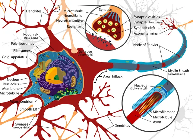
A neuron combines a cell body, dendrites that receive signals, and a single axon that conducts the message toward other cells. Its functional polarity imposes a direction on the flow of information.
It is a cell with very high energy needs and, in almost all regions, unable to divide in adults: its loss is, as a rule, permanent.

Glia
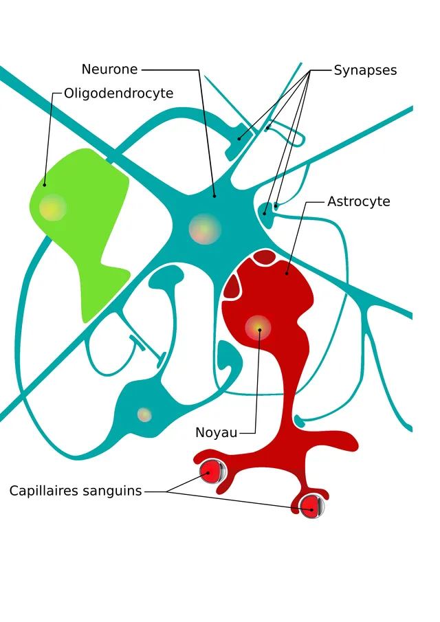
Glial cells are at least as numerous as neurons — the ratio, long put at ten to one, is probably closer to one to one in the human brain. They are not mere support cells.

The neuron and its processes
Most of the neuron's proteins are synthesized in the cell body: what the axon needs has to be shipped there. This axonal transport, anterograde by kinesin and retrograde by dynein, covers up to several hundred millimeters a day. Some viruses, including rabies and herpes, use it to travel up to the central nervous system.
This dependence explains chromatolysis: after an axon is cut, the cell body swells and its Nissl bodies disperse, a sign of metabolic reorganization toward repair.
Glial cells, the majority and indispensable
The astrocyte provides the neuron's metabolic support. Its perivascular end-feet surround the capillaries and contribute to the blood-brain barrier; it takes back glutamate released into the synaptic cleft, whose accumulation would be toxic; it buffers extracellular potassium.
The oligodendrocyte myelinates the axons of the central nervous system, each one able to wrap several dozen. In the periphery, the Schwann cell does the job, but one cell for a single segment of a single axon. After injury, Schwann cells form a guide tube and an environment that favors regrowth — hence the much better regeneration of peripheral nerves.
Microglia, finally, are the resident macrophages of the nervous system. They come from the yolk sac and not from the neuroectoderm, which makes them the only glial cells of different origin.
The shapes of the neuron
Neurons are classified by the number of processes leaving the cell body. The multipolar neuron, by far the most common, has many dendrites and one axon: this applies to motor neurons and most neurons of the brain. The bipolar neuron has one dendrite and one axon on opposite sides: it is found in the retina and the inner ear. The pseudo-unipolar neuron has a single process that divides in a T: it is the sensory neuron of the spinal ganglia, one branch going to the skin and the other to the spinal cord.
They are also classified by function: sensory neurons, which bring information in; motor neurons, which command muscles; and interneurons, by far the most numerous, which link neurons together.
The cell body contains Nissl bodies, clumps of rough reticulum, extending into the large dendrites but not into the axon hillock or the axon itself. The neuronal cytoskeleton comprises microtubules, the rails of axonal transport, and neurofilaments, intermediate filaments that set the calibre of the axon.
Myelin and its diseases
Myelin is a tight wrapping of plasma membrane around the axon, very rich in lipids. In the central nervous system, one oligodendrocyte myelinates a segment on several dozen axons; in the periphery, one Schwann cell myelinates only one segment of a single axon. Between two segments, the nodes of Ranvier concentrate the sodium channels.
Not all fibres are myelinated. In the periphery, Schwann cells house several small axons in folds of their membrane without wrapping them in myelin: these are unmyelinated fibres, slow, which carry dull pain and the commands of the autonomic nervous system, among others.
In multiple sclerosis, the immune system destroys central myelin in patches: conduction slows or stops, causing relapses of visual, motor or sensory disturbance. Guillain-Barré syndrome attacks peripheral myelin, often after an infection, and causes paralysis that rises from the legs to the trunk. The Schwann cell can make new myelin, which explains why recovery is the rule in the latter.
The peripheral nerve and its repair
A nerve is wrapped like a muscle. The epineurium, dense connective tissue, surrounds the whole nerve; the perineurium surrounds each fascicle and, through its tightly joined cells, forms a barrier that protects the axons' environment; the endoneurium surrounds each fibre.
After an axon is cut, the segment separated from the cell body degenerates: this is Wallerian degeneration. Macrophages and Schwann cells clear the debris, then the Schwann cells line up in columns that will serve as guides. The proximal end of the axon sprouts and regrows along these columns at a rate of about one millimetre per day.
Surgical repair consists of bringing the two ends of the nerve together so that the axons find their sheaths again. The further the injury is from the muscle, the longer recovery takes: an axon cut in the arm takes several months to reach the hand.
The blood-brain barrier and cerebrospinal fluid
Brain capillaries differ from those of other organs. Their endothelial cells are sealed by continuous tight junctions, have almost no transport vesicles and are surrounded by pericytes and astrocyte end-feet. Only small lipid-soluble molecules pass freely, such as oxygen, carbon dioxide, alcohol or anaesthetics; glucose and amino acids use transporters.
This barrier protects neurons but hampers treatment. Dopamine does not cross it: its precursor, levodopa, is given instead, which uses an amino-acid transporter and is converted into dopamine in the brain. A few areas, the circumventricular organs, lack the barrier, so that the brain can monitor blood composition or release hormones into it.
Cerebrospinal fluid is secreted by the choroid plexuses of the ventricles, made of a cuboidal epithelium around capillaries. It flows from the ventricles into the spaces surrounding the brain and spinal cord, then joins the venous blood. It cushions the brain, which floats in it, and sampling it by lumbar puncture helps diagnose meningitis.
Glia in the face of injury
When the central nervous system is injured, microglia activate first: they move to the lesion, phagocytose debris and release inflammatory signals. Astrocytes proliferate and enlarge, forming a glial scar that isolates the area but also hinders axon regrowth.
Primary brain tumours almost all arise from glial cells, which can divide, and not from adult neurons, which no longer divide. Glioblastoma, the most aggressive astrocytic tumour, is the most common primary malignant brain tumour in adults; it infiltrates the neighbouring tissue, which makes complete removal impossible. In adults, however, the most common tumours in the brain are metastases from cancers elsewhere.
58 answered questions go with this chapter in the practice section.
Sources and further reading
Reference textbooks
- Alan Stevens, James Lowe, Histologie humaine, Elsevier Masson.
Open access
- 4.5 Nervous Tissue Mediates Perception and Response — OpenStax, Anatomy and Physiology 2e.
Last revised : . Text licensed under CC BY-SA 4.0.