Muscle tissues
Skeletal striated, cardiac striated and smooth muscle.
Three varieties, three kinds of control
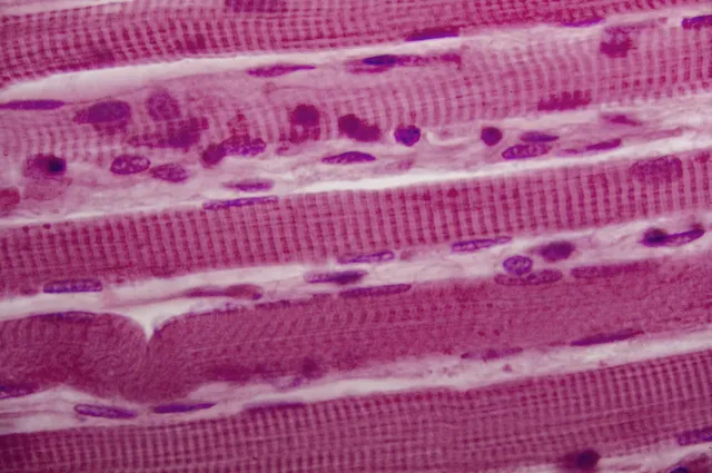
Skeletal striated muscle is made of long multinucleated fibers, with peripheral nuclei, under voluntary control.
Cardiac striated muscle is made of short cells with one or two central nuclei, linked by intercalated discs and under involuntary control. Its automaticity is intrinsic.
Smooth muscle is made of spindle-shaped cells with a single central nucleus, without striations, under involuntary control. It is found in the walls of vessels and hollow viscera.

The sarcomere, the unit of contraction
In striated muscle, actin and myosin are arranged in repeating units, the sarcomeres, which stretch from one Z line to the next. This regular alignment produces the striation visible under the microscope.
The A band, dark, corresponds to the length of the thick myosin filaments. The I band, light, contains only thin actin filaments. During contraction, the filaments do not shorten: they slide over one another. The A band therefore keeps its length, while the I band and the whole sarcomere shorten.
The force developed depends on the number of possible cross-bridges between myosin and actin, and therefore on the overlap between the two sets of filaments. Overstretched, the sarcomere no longer has enough overlap; overshortened, the filaments get in each other's way. There is an optimal length, and it is the mechanical basis of the Frank-Starling law in the heart.
Smooth muscle, a different organization
Smooth muscle contains the same contractile proteins, but arranged obliquely and anchored to dense bodies rather than aligned in sarcomeres. Hence the absence of striation, and a contraction that is slow but very economical: it can hold tone for hours at a negligible energy cost.
Its control differs too. Calcium acts there not through troponin, which is absent, but through calmodulin, which activates myosin light-chain kinase. This slower mechanism explains the delayed response.
Finally, many smooth muscle fibers are coupled by gap junctions and contract as a sheet, sometimes with their own automaticity — intestinal peristalsis persists in a segment cut off from all innervation.
From muscle to myofibril
A skeletal muscle is wrapped at three levels. The epimysium, dense connective tissue, surrounds the whole muscle; the perimysium divides it into bundles of fibres; the very thin endomysium surrounds each fibre and carries the capillaries. These sheaths continue into the tendon and transmit force to the bone.
The muscle fibre is a giant cell, formed by the fusion of many myoblasts, hence its hundreds of nuclei pressed beneath the membrane. Its cytoplasm is packed with myofibrils, cylinders of sarcomeres lined up end to end, and wrapped in a sarcoplasmic reticulum that stores calcium.
The membrane dips into the fibre as transverse tubules, the T tubules. Each T tubule is flanked by two cisterns of the reticulum: together they form a triad. When the action potential runs down the tubule, a voltage sensor in the membrane changes shape and mechanically opens the reticulum's calcium channels, the ryanodine receptors. Calcium floods the cytoplasm, binds to troponin and frees the binding sites on actin.
Fibre types and recruitment
Not all fibres are equal. Type I fibres, slow, rich in mitochondria, capillaries and myoglobin, hence their red colour, work aerobically and resist fatigue: they dominate in postural muscles. Type IIx fibres, fast, poor in mitochondria and rich in glycogen, produce high force through glycolysis but tire quickly. IIa fibres are intermediate.
All the fibres of one motor unit are of the same type, because the motor neuron imposes their properties. During increasing effort, units are recruited in a fixed order, from the smallest, slow and resistant, to the largest, fast and powerful: this is the size principle. A fine movement therefore mainly uses small units, and force can be graded precisely.
Training modifies fibres more than it creates them. Endurance increases mitochondria and capillaries; strength training increases fibre diameter by adding myofibrils, helped by satellite cells that supply new nuclei.
When skeletal muscle is diseased
Duchenne muscular dystrophy is due to the absence of dystrophin, a large protein that links the fibre's cytoskeleton to the extracellular matrix. Without it, the membrane tears with each contraction and the fibres die. The gene lies on the X chromosome: the disease affects almost only boys, begins in childhood with falls and difficulty getting up, and comes with a massive rise in blood creatine kinase.
Myasthenia gravis is an autoimmune disease of the neuromuscular junction: antibodies block or destroy acetylcholine receptors. Muscle tires abnormally with repeated effort, typically with drooping eyelids at the end of the day and double vision. Drugs that inhibit the breakdown of acetylcholine improve it.
Rhabdomyolysis is a massive destruction of fibres, after crushing, extreme exertion or certain drugs. The released myoglobin passes into the urine, turning it brown, and can block the kidney tubules.
The cardiac muscle cell in detail
The cardiac muscle cell is striated like the skeletal fibre, but short and branched, with one or two central nuclei. Its intercalated discs combine three junctions: adherens junctions and desmosomes, which pass traction from one cell to the next, and gap junctions, which let current through. The myocardium thus behaves as a continuous electrical network.
Its coupling differs from that of skeletal muscle. The action potential opens calcium channels in the membrane; this small influx of extracellular calcium triggers the opening of the reticulum's channels, which release the rest. Without external calcium the heart stops contracting, whereas skeletal muscle keeps going for a while.
The cardiac muscle cell is one of the richest in mitochondria, which occupy about a third of its volume: the heart never stops and depends almost entirely on aerobic metabolism. Deprived of oxygen, it dies within a few tens of minutes and releases its proteins, including cardiac troponin, measured in the blood to diagnose a heart attack.
The many faces of smooth muscle
Smooth muscle comes in two forms. In single-unit smooth muscle, the most widespread, cells are coupled by gap junctions and contract together: the wall of the digestive tract, the uterus, the bladder. In multi-unit smooth muscle, each cell is controlled by its own nerve ending, for fine adjustment: the muscles of the iris, the muscles that raise the hairs.
Its contraction is slow to develop but can be maintained for a very long time with little ATP, thanks to cross-bridges that stay attached. This allows arteries to keep a permanent tone and sphincters to stay closed.
Unlike striated muscle cells, the smooth muscle cell can divide. The uterus in pregnancy enlarges through both an increase in the size of its cells and in their number. Close relatives of smooth muscle, myoepithelial cells surround the acini of glands and squeeze out their secretion, and pericytes surround capillaries.
58 answered questions go with this chapter in the practice section.
Sources and further reading
Reference textbooks
- Alan Stevens, James Lowe, Histologie humaine, Elsevier Masson.
Open access
- 4.4 Muscle Tissue and Motion — OpenStax, Anatomy and Physiology 2e.
Last revised : . Text licensed under CC BY-SA 4.0.