Cardiovascular physiology
Automaticity, cardiac output, blood pressure and its regulation.
Cardiac automaticity
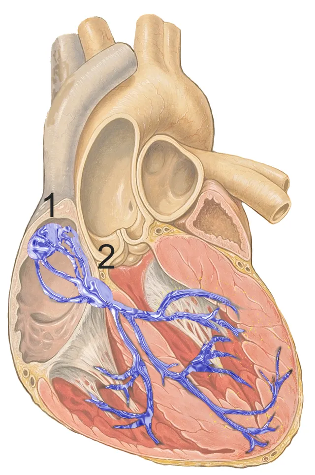
The heart beats without nervous command: the cells of the sinoatrial node depolarize spontaneously and set the rhythm. The autonomic nervous system only modulates this intrinsic rate.
The sympathetic system speeds it up and strengthens contraction; the parasympathetic system, through the vagus nerve, slows it down. At rest, vagal tone predominates — hence a rate well below that of the isolated sinoatrial node.

Output and pressure
Cardiac output is the product of heart rate and stroke volume. Mean arterial pressure, in turn, results from the product of cardiac output and total peripheral resistance.
This resistance is adjusted almost entirely by the arterioles, whose caliber varies. Poiseuille's law explains why: resistance varies inversely with the fourth power of the radius.
The cardiac cycle, phase by phase
Systole begins with isovolumetric contraction: the atrioventricular valves close, the semilunar valves are not yet open, the volume does not change but the pressure rises quickly. When it exceeds aortic pressure, the semilunar valves open and ejection begins.
Diastole likewise begins with isovolumetric relaxation, the semilunar valves having closed, until ventricular pressure falls below atrial pressure. Filling then happens mostly passively; atrial contraction only adds about twenty percent, but that share becomes critical when the heart is stiff or fast.
The valves never open or close actively: they follow pressure gradients. The chordae and papillary muscles only stop them from turning inside out.
Two cardiac action potentials
The action potential of a ventricular cardiac muscle cell lasts about 250 to 300 milliseconds, a hundred times longer than that of a neuron. After a rapid depolarisation due to sodium, it holds on a long plateau, where calcium entry balances potassium exit, before repolarisation through potassium channels. This plateau extends the refractory period almost to the end of contraction: the heart cannot go into tetanus, and it always relaxes between beats, which allows it to fill.
Cells of the sinus node have no stable resting potential. After each action potential, their membrane slowly depolarises on its own, notably thanks to a special current activated by hyperpolarisation, until it reaches threshold. Their rapid depolarisation is due to calcium, not sodium, hence its relative slowness.
Heart rate depends on the slope of this slow depolarisation. The sympathetic system, through beta-1 receptors, steepens it and speeds the heart; the vagus nerve, through acetylcholine, flattens it and hyperpolarises the cell, which slows the heart. Beta-blockers slow the heart by blocking the sympathetic effect; another drug does so by directly blocking the current of slow depolarisation.
Reading an electrocardiogram
The electrocardiogram records the heart's electrical activity at the body surface. The P wave reflects depolarisation of the atria. The PR interval, 0.12 to 0.20 seconds, measures mainly passage through the atrioventricular node, which deliberately delays the wave to let the atria finish filling the ventricles. The QRS complex, brief, under 0.12 seconds, reflects depolarisation of the ventricles; the T wave, their repolarisation. Atrial repolarisation is hidden by the QRS.
The paper runs at 25 millimetres per second: a large 5 mm square is worth 0.2 seconds. Heart rate is estimated by dividing 300 by the number of large squares between two QRS complexes: one QRS every four large squares corresponds to 75 beats per minute.
Some abnormalities can be read directly. A prolonged PR signals slowed atrioventricular conduction; P waves not followed by QRS, a block. In atrial fibrillation, a common rhythm disorder in older people, P waves disappear and QRS complexes occur completely irregularly. A wide QRS suggests abnormal conduction within the ventricles.
Pressure, resistance and calibre
Mean arterial pressure is the product of cardiac output and total peripheral resistance, which lies mainly in the arterioles. As diastole lasts longer than systole, mean pressure is closer to diastolic pressure: it is estimated as diastolic plus one third of the difference. For 120/80 mmHg, it is about 93 mmHg.
The resistance of a vessel varies inversely with the fourth power of its radius: if the radius halves, resistance is multiplied by sixteen. This is why slight constriction of arterioles is enough to redistribute flow between organs, and why moderate narrowing of a coronary artery can cause symptoms during exertion.
Pulse pressure, the difference between systolic and diastolic, depends on the volume ejected and the stiffness of the large arteries. With age, the aorta loses its elasticity: systolic pressure rises, diastolic falls, and pulse pressure widens.
Slow adjustments and standing up
The baroreflex corrects pressure within seconds but does not regulate it over the long term. That role falls to the kidney, which adjusts blood volume: when pressure rises, it excretes more sodium and water; when it falls, the renin-angiotensin-aldosterone system and antidiuretic hormone retain sodium and water. Conversely, atria stretched by excess volume secrete a natriuretic peptide that promotes sodium excretion.
On moving from lying to standing, about half a litre of blood pools in the leg veins. Venous return and cardiac output fall, pressure drops for a moment; the baroreflex immediately speeds the heart and constricts the vessels.
When this correction fails, for example in older people, in dehydration or with certain drugs, pressure stays low: this is orthostatic hypotension, defined as a fall of at least 20 mmHg in systolic or 10 mmHg in diastolic pressure within three minutes of standing. It causes dizziness and falls.
Special circulations
The myocardium already extracts about 70% of the oxygen from coronary blood at rest: to receive more oxygen during exertion, it can only increase coronary flow. The left ventricle, whose wall compresses the arteries during systole, is perfused mainly in diastole; a fast heart rate, which shortens diastole, therefore reduces its perfusion time.
The brain keeps its blood flow constant over a wide range of arterial pressure through autoregulation. It is very sensitive to carbon dioxide: hyperventilation, by lowering CO2, constricts cerebral arteries and can cause dizziness.
The pulmonary circulation works at low pressure, about six times lower than the systemic circulation. Its arterioles constrict where alveolar oxygen is lacking, which directs blood towards well-ventilated areas; extended to the whole lung, at altitude or in some respiratory diseases, this response raises pulmonary pressure. The skin circulation, finally, serves mainly to regulate temperature.
58 answered questions go with this chapter in the practice section.
Sources and further reading
Reference textbooks
- John E. Hall, Michael E. Hall, Guyton et Hall. Physiologie médicale, Elsevier Masson.
Open access
- 19.2 Cardiac Muscle and Electrical Activity — OpenStax, Anatomy and Physiology 2e.
- 19.3 Cardiac Cycle — OpenStax, Anatomy and Physiology 2e.
Last revised : . Text licensed under CC BY-SA 4.0.