Resting potential and action potential
Ion gradients, threshold, depolarization and propagation.
Where the resting potential comes from
At rest, the membrane is far more permeable to potassium than to sodium. Potassium, abundant inside, tends to leave down its chemical gradient, but every ion that leaves behind a negative charge that pulls it back. An equilibrium sets in, around minus ninety millivolts for potassium alone.
The real potential, around minus seventy millivolts, is a little less negative because a small sodium permeability pulls the other way. The resting potential is therefore an average weighted by permeabilities.
The sodium-potassium pump is not the direct source of this potential: it maintains the gradients on which it rests. Its own electrogenic contribution, due to the three-to-two ratio, does not exceed a few millivolts.
All or nothing
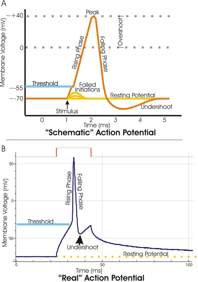
Once the threshold is reached, voltage-gated sodium channels open massively and depolarization becomes explosive. Their inactivation, followed by the delayed opening of potassium channels, brings the potential back to its resting value.
The amplitude of the action potential does not depend on the intensity of the stimulus: a stronger stimulus increases the frequency of action potentials, not their size.

Saltatory conduction
On a myelinated axon, the sheath insulates the membrane except at the nodes of Ranvier. The action potential is only regenerated at these nodes and seems to jump from one to the next.
There is a double gain: speed increases considerably, and energy expenditure decreases since ion fluxes only involve a fraction of the surface.
Nernst and Goldman
The equilibrium potential of an ion is the voltage at which the electrical force exactly balances its concentration gradient: the ion has no net flux. The Nernst equation calculates it; at 37 °C, for a monovalent ion, it equals about 61 mV times the base-ten logarithm of the ratio of outside to inside concentrations.
For potassium, 4 mmol/L outside and 140 inside, the ratio is about 1/35, whose logarithm is about −1.5: the equilibrium potential is about −90 mV. For sodium, 140 outside and 15 inside, it is about +60 mV. For chloride, an anion, the sign reverses and it lies around −70 mV.
When the membrane is permeable to several ions, the potential settles between their equilibrium potentials, the closer to one of them the more permeable the membrane is to it: this is what the Goldman equation formalises. At rest, dominated by potassium, it is close to −70 mV; at the peak of the action potential, dominated by sodium, it approaches +60 mV without reaching it.
The channels of the action potential
The voltage-gated sodium channel has two gates. The activation gate, closed at rest, opens very quickly when the membrane depolarises to threshold, around −55 mV. The inactivation gate closes a little more slowly, in less than a millisecond, and plugs the channel even if depolarisation persists. The channel can open again only after the membrane has returned towards rest.
The voltage-gated potassium channel opens with a delay. Its opening lets potassium out and brings the potential back towards rest; as it closes slowly, the potential dips briefly below the resting value: this is the after-hyperpolarisation. The whole event lasts about one millisecond in a neuron.
The amounts of ions crossing the membrane during an action potential are tiny compared with the stores: a single potential does not change concentrations measurably. The sodium-potassium pump therefore plays no part in each action potential, but acts over time to maintain the gradients.
Coding an intensity
Since the action potential always has the same amplitude, stimulus intensity is coded in other ways. First by frequency: a stronger stimulus brings the membrane back to threshold faster after each potential, during the relative refractory period, and potentials follow each other more closely. Second by recruitment: a stronger stimulus activates more fibres.
The absolute refractory period, due to inactivation of sodium channels, sets a maximum frequency, of the order of a few hundred potentials per second. It also imposes the direction of propagation: the region just excited cannot be excited again straight away, and the wave can only move forward.
Nerve fibres and their speed
Conduction velocity depends on axon diameter and myelination. Large myelinated Aα fibres, which command muscles and carry proprioception, conduct at 70 to 120 metres per second. Aβ fibres, slightly thinner, carry touch. Aδ fibres, thin and lightly myelinated, conduct at 5 to 30 m/s. C fibres, unmyelinated, do not exceed 2 m/s.
The difference can be felt when a finger is struck: a first sharp, well-localised pain, carried by Aδ fibres, comes nearly a second before a dull, diffuse pain, carried by C fibres.
Nerve conduction studies measure the conduction velocity of nerves. A very slow velocity suggests myelin damage; reduced amplitude with preserved velocity suggests loss of axons.
Drugs and poisons
Tetrodotoxin, the poison of the puffer fish, plugs voltage-gated sodium channels: action potentials no longer arise, and paralysis can reach the respiratory muscles. Local anaesthetics, such as lidocaine, block the same channels, but reversibly and locally.
Local anaesthetics reach thin fibres faster than thick ones: pain disappears first, then temperature sensation and touch, and movement last. They act more on channels that open often, and therefore on the most active fibres. The same principle explains the use of sodium channel blockers against some epilepsies and some heart rhythm disorders.
Extracellular ions also alter excitability. Hyperkalaemia depolarises the membrane and, if prolonged, inactivates sodium channels; hypokalaemia hyperpolarises it and causes muscle weakness and arrhythmias. Hypocalcaemia lowers the threshold and makes nerves and muscles hyperexcitable: tingling, cramps, even spasms, known as tetany.
58 answered questions go with this chapter in the practice section.
Sources and further reading
Reference textbooks
- John E. Hall, Michael E. Hall, Guyton et Hall. Physiologie médicale, Elsevier Masson.
Open access
- 12.4 The Action Potential — OpenStax, Anatomy and Physiology 2e.
Last revised : . Text licensed under CC BY-SA 4.0.