Synaptic transmission
Chemical synapse, neurotransmitters, summation and neuromuscular junction.
The chemical relay
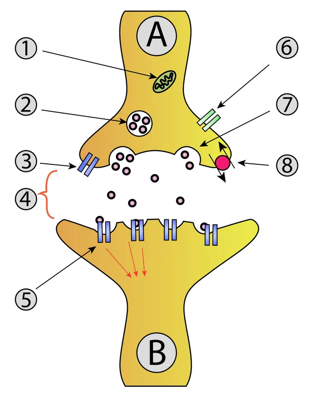
When the action potential reaches the terminal, it opens voltage-gated calcium channels. Calcium entry triggers vesicle fusion and the release of the neurotransmitter into the cleft.
Calcium is therefore the real messenger of excitation-secretion coupling: without calcium entry, there is no release, whatever the depolarization.

Postsynaptic integration
The neurotransmitter generates an excitatory or inhibitory postsynaptic potential depending on the receptor — and therefore on the target cell, not on the transmitter itself.
These graded potentials add up over time and space. The neuron only fires an action potential if their sum reaches threshold at the initial segment of the axon: that is where the decision is made.
The complete path of a signal
The transmitter diffuses across the cleft, about twenty nanometers wide, and binds to postsynaptic receptors. If they are ionotropic, the channel opens at once and the effect is immediate but brief. If they are metabotropic, an intracellular cascade is set in motion: a slower, longer-lasting, but amplified effect.
Finally, the signal must be switched off, otherwise the receptor would stay stimulated permanently. Three routes handle this, depending on the transmitter: enzymatic breakdown in the cleft, reuptake by the neuron or by the astrocyte, and simple diffusion out of the cleft.
The life of a vesicle
The neurotransmitter is made in the terminal, or arrives there by axonal transport, then concentrated into vesicles by a vesicular transporter. Some vesicles are docked at the membrane, ready to fuse, in active zones rich in calcium channels.
Fusion relies on SNARE proteins, carried on one side by the vesicle and on the other by the membrane, which wind around each other like a zip. A calcium sensor, synaptotagmin, triggers fusion when calcium enters: release occurs less than a millisecond after the action potential arrives. The vesicle membrane is then retrieved by endocytosis and recycled.
Two bacterial toxins cut SNARE proteins. Botulinum toxin acts at the neuromuscular junction: acetylcholine is no longer released and paralysis is flaccid. It is used in very small doses to relax overactive muscles. Tetanus toxin, by contrast, travels to the spinal cord and blocks release in inhibitory interneurons: with their brakes removed, motor neurons run wild and muscles contract continuously.
Two families of receptors
Ionotropic receptors are themselves channels: binding of the neurotransmitter opens a pore and ions pass through within a fraction of a millisecond. The nicotinic acetylcholine receptor, the AMPA and NMDA glutamate receptors and the GABA-A receptor belong to this group. They provide fast, point-to-point transmission.
Metabotropic receptors, coupled to G proteins, have no pore. They activate an intracellular cascade, for example production of cAMP, which modifies channels or gene expression. Their effect is slower, from tens of milliseconds to several minutes, but amplified and widespread. Muscarinic acetylcholine receptors, adrenergic and dopaminergic receptors and most serotonin receptors are metabotropic.
The same neurotransmitter can thus have opposite effects depending on the receptor: acetylcholine contracts skeletal muscle through a nicotinic receptor, but slows the heart through a muscarinic receptor.
Stopping the signal, and how drugs use it
A synaptic signal makes sense only if it is brief. Three mechanisms stop it. Acetylcholine is broken down on the spot by acetylcholinesterase. Monoamines — dopamine, noradrenaline, serotonin — are taken back up by transporters in the presynaptic terminal. Glutamate is largely taken up by neighbouring astrocytes. Some simply diffuses out of the cleft.
Many drugs, medical and recreational, act on these steps. The most prescribed antidepressants block serotonin reuptake and prolong its action. Cocaine blocks reuptake of dopamine and noradrenaline. Anticholinesterases slow the breakdown of acetylcholine: they are used in myasthenia gravis and in Alzheimer's disease.
Organophosphate insecticides and some nerve agents inhibit acetylcholinesterase irreversibly. Acetylcholine builds up everywhere: sweating, salivation, tearing, congested airways, diarrhoea, pinpoint pupils, slow heart, then muscle paralysis. Atropine, which blocks muscarinic receptors, corrects some of these signs.
The major neurotransmitter systems
Glutamate is the main excitatory neurotransmitter of the central nervous system; GABA is the main inhibitory one in the brain, and glycine in the spinal cord. Strychnine, which blocks glycine receptors, causes convulsions. Benzodiazepines strengthen the effect of GABA on its receptor, hence their calming and anticonvulsant action.
Monoamines come from small groups of brainstem neurons whose axons branch throughout the brain. Dopamine neurons of the substantia nigra project to the striatum to control movement: their death causes Parkinson's disease. Others, in the ventral tegmental area, take part in the reward circuit, the target of addictive drugs. Serotonin comes from the raphe nuclei, noradrenaline from the locus coeruleus; both modulate mood, alertness and sleep.
Acetylcholine in the central nervous system, coming notably from a nucleus at the base of the brain, contributes to attention and memory; these neurons degenerate early in Alzheimer's disease. Neuropeptides, such as endorphins, are often released together with a classic neurotransmitter and modulate its effect.
The NMDA receptor, a coincidence detector
The NMDA glutamate receptor has a peculiarity: at rest, its channel is plugged by a magnesium ion. It opens only if two conditions are met at the same time: glutamate bound, and a membrane already depolarised, which expels the magnesium. It therefore detects coincidence between activity of the presynaptic and postsynaptic neurons.
Once open, it lets calcium in, which activates enzymes and strengthens the synapse lastingly, for example by adding AMPA receptors: this is long-term potentiation, studied in the hippocampus as a cellular basis of memory.
The same mechanism becomes toxic in a stroke. Deprived of energy, neurons release massive amounts of glutamate that astrocytes no longer take up; excessive calcium entry through NMDA receptors kills neighbouring neurons: this is excitotoxicity.
58 answered questions go with this chapter in the practice section.
Sources and further reading
Reference textbooks
- John E. Hall, Michael E. Hall, Guyton et Hall. Physiologie médicale, Elsevier Masson.
Open access
- 12.5 Communication Between Neurons — OpenStax, Anatomy and Physiology 2e.
Last revised : . Text licensed under CC BY-SA 4.0.