Respiratory physiology
Volumes, alveolar exchange, gas transport and control.
Ventilating is not exchanging
Ventilation renews alveolar air; diffusion moves gases between alveolus and capillary; perfusion carries them away. All three must be matched: an alveolus that is ventilated but not perfused is useless, and so is the reverse.
Dead space is the volume that is ventilated without exchanging — the conducting airways. This is why rapid, shallow breathing ventilates poorly, despite an identical total airflow.

Gas transport
Oxygen is carried almost entirely bound to hemoglobin; the dissolved fraction is negligible. The dissociation curve is sigmoid, reflecting cooperativity between the four subunits.
Carbon dioxide travels three ways: dissolved, bound to hemoglobin, and above all as bicarbonate — which closely links respiration and acid-base balance.
Ventilation, diffusion, perfusion
Three steps separate ambient air from arterial blood, and each can fail independently.
Ventilation brings air to the alveoli. About one hundred and fifty milliliters of each breath stays in the conducting airways without taking part in exchange: this is the anatomical dead space. Rapid, shallow breathing moves a lot of air for very little useful alveolar ventilation.
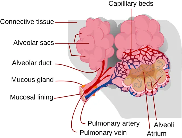
Diffusion carries gases across the alveolar-capillary barrier, less than one micrometer thick over a surface of around seventy square meters. Carbon dioxide diffuses about twenty times better than oxygen, so a diffusion defect first shows up as isolated hypoxemia.
Perfusion, finally, must match the ventilated alveoli. An alveolus that is ventilated but not perfused is dead space; one that is perfused but not ventilated is a shunt, and oxygen does not correct the latter.
The mechanics of ventilation
Inspiration is always active. The diaphragm, by contracting, descends and enlarges the thorax from top to bottom; the external intercostal muscles lift the ribs. Thoracic volume increases, pressure in the alveoli falls slightly below atmospheric pressure, and air flows in. At rest, expiration is passive: the elastic lung recoils as soon as the muscles relax. It becomes active only during exertion or in certain diseases, using the abdominal muscles.
The lung tends to recoil inwards, the chest wall to spring outwards: between them, the pleural cavity keeps a negative pressure, about −5 cmH2O at rest, which holds the lung against the wall. If air enters the pleura, through a breach in the wall or the lung, this pressure disappears and the lung collapses: this is pneumothorax.
Resistance to airflow lies mainly in the medium-sized bronchi, not in the smallest ones, which are very numerous and form a large total cross-section. In asthma, contraction of bronchial smooth muscle, inflammation and mucus narrow these bronchi, and resistance rises sharply, especially during expiration.
Lung volumes and spirometry
In an adult at rest, each breath moves about 500 mL: this is the tidal volume. Beyond a normal inspiration, one can still inhale about 3 litres, the inspiratory reserve volume; beyond a normal expiration, still exhale about 1.2 litres, the expiratory reserve volume. About 1.2 litres of air always remains in the lungs, the residual volume, which no expiration can expel.
Capacities are sums of volumes. Vital capacity combines tidal volume and the reserve volumes; total lung capacity adds the residual volume; functional residual capacity, the volume present at the end of a quiet expiration, combines expiratory reserve volume and residual volume. Residual volume, and hence these last two capacities, cannot be measured by simple spirometry: plethysmography or a gas dilution technique is needed.
Spirometry also measures the maximum volume exhaled in the first second, the FEV1. Divided by vital capacity, it defines the Tiffeneau ratio: below 0.7, it indicates an obstructive ventilatory defect, as in asthma or chronic obstructive pulmonary disease. A restrictive defect, by contrast, is defined by a reduced total lung capacity, with a preserved ratio.
- Tidal volume: about 0.5 L.
- Inspiratory reserve volume: about 3 L.
- Expiratory reserve volume: about 1.2 L.
- Residual volume: about 1.2 L.
Exchange across the alveolus
In the alveolus, the partial pressure of oxygen is about 100 mmHg and that of carbon dioxide about 40 mmHg. Venous blood arriving has an oxygen pressure of about 40 mmHg and a CO2 pressure of about 46 mmHg: each gas diffuses towards the side where its pressure is lower. Arterial blood leaves with about 95 mmHg of oxygen and 40 mmHg of CO2.
Diffusion follows Fick's law: gas flow is proportional to the exchange surface and the pressure difference, and inversely proportional to membrane thickness. Emphysema reduces the surface, fibrosis thickens the membrane: both impair diffusion. CO2, much more soluble, diffuses about twenty times more easily than oxygen; this is why diffusion defects lower oxygen long before they raise CO2.
At altitude, the proportion of oxygen in air remains 21%, but atmospheric pressure falls: the partial pressure of inspired oxygen falls accordingly. At 5,000 metres, it is reduced to about half its sea-level value.
Blood gases
Arterial blood gas analysis measures pH, the partial pressure of oxygen, that of carbon dioxide, and bicarbonate. In a healthy adult, pH lies between 7.38 and 7.42, PaCO2 between 35 and 45 mmHg, bicarbonate between 22 and 26 mmol/L.
The lung regulates CO2, and therefore the respiratory part of acid-base balance. Hypoventilation accumulates CO2 and causes respiratory acidosis; hyperventilation, from anxiety or at altitude, lowers CO2 and causes respiratory alkalosis. The kidney compensates slowly, over a few days, by retaining or excreting bicarbonate.
Hypoxaemia, a fall in arterial oxygen, has several causes: hypoventilation, which also raises CO2; poorly ventilated but perfused areas, the most common cause; a shunt, where blood crosses the lung without meeting air, and which administered oxygen corrects poorly; a diffusion defect; and altitude.
Altitude and carbon monoxide
At altitude, the fall in oxygen stimulates peripheral chemoreceptors and causes hyperventilation: CO2 falls, causing alkalosis, which the kidney compensates within a few days. Over weeks, erythropoietin increases the number of red cells. Ascending too fast can cause acute mountain sickness — headache, nausea, insomnia — and, more rarely, pulmonary or cerebral oedema, which require descent.
Carbon monoxide binds to haemoglobin with an affinity about two hundred times greater than that of oxygen. It takes the place of oxygen and also hinders its release in the tissues. The partial pressure of oxygen in the blood stays normal, and the usual pulse oximeter, which confuses the two forms, displays a falsely normal saturation: diagnosis relies on measuring carboxyhaemoglobin.
Treatment consists of giving high-flow oxygen, which speeds elimination of carbon monoxide, or even pressurised oxygen in a hyperbaric chamber. Poorly maintained heaters and braziers in enclosed spaces are the usual causes, often in winter and affecting a whole family.
58 answered questions go with this chapter in the practice section.
Sources and further reading
Reference textbooks
- John E. Hall, Michael E. Hall, Guyton et Hall. Physiologie médicale, Elsevier Masson.
Open access
- 22.4 Gas Exchange — OpenStax, Anatomy and Physiology 2e.
- 22.5 Transport of Gases — OpenStax, Anatomy and Physiology 2e.
Last revised : . Text licensed under CC BY-SA 4.0.