Abdomen: peritoneum, regions and digestive organs
The peritoneal membrane, position of the viscera, stomach and intestine.
The stomach
The stomach churns the food bolus and begins protein digestion. Its mucosa secretes hydrochloric acid and pepsinogen, and protects itself with an alkaline mucus.
Its muscularis has a third, inner oblique layer, absent from the rest of the digestive tract: it reinforces the churning that turns the food bolus into chyme.

The colonic frame
The colon frames the loops of small bowel: ascending on the right, transverse at the top, descending on the left, then sigmoid before the rectum. The ascending and descending colon are fixed to the posterior wall; the transverse and sigmoid colon, suspended by a mesentery, are mobile.
Its main function is to reabsorb water and electrolytes. Its longitudinal muscle, gathered into three bands (taeniae coli), puckers the wall into characteristic haustra.
The peritoneum, and the geography it imposes
The peritoneum is a two-layered serous membrane: parietal against the wall, visceral around the organs. Between the two lies a potential cavity. The folds linking the two layers — mesenteries, omenta, ligaments — carry vessels and nerves to the organs.
An organ suspended by a mesentery is intraperitoneal and mobile: stomach, jejunum, ileum, transverse and sigmoid colon. An organ pressed against the posterior wall is retroperitoneal and fixed: kidneys, adrenal glands, most of the pancreas, aorta, vena cava, ascending and descending colon.
This distinction governs the surgical approach, the spread of infection and the interpretation of imaging.
Dividing the abdomen into regions
To describe a symptom or examination finding, the front of the abdomen is divided by two vertical lines, the midclavicular lines, and two horizontal lines: one below the costal margin, the other through the tubercles of the iliac crests. This gives nine regions.
The upper row comprises the right hypochondrium, the epigastrium and the left hypochondrium; the middle row, the right flank, the umbilical region and the left flank; the lower row, the right iliac fossa, the hypogastrium and the left iliac fossa. In emergencies, four quadrants defined by two lines crossing at the umbilicus are often enough.
Each region has its usual organs: liver and gallbladder in the right hypochondrium, stomach in the epigastrium, spleen in the left hypochondrium, caecum and appendix in the right iliac fossa, sigmoid colon in the left iliac fossa, full bladder in the hypogastrium.
The stomach and its folds
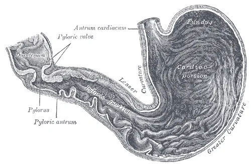
The stomach is divided into the cardia, where the oesophagus enters, the fundus, a dome above the cardia often filled with air, the body, the antrum, then the pylorus, a sphincter opening into the duodenum. Its concave right border is the lesser curvature; its convex left border, the greater curvature.
Two peritoneal folds attach to it. The lesser omentum links the lesser curvature and the start of the duodenum to the liver; its free edge, the hepatoduodenal ligament, contains the hepatic pedicle. The greater omentum hangs from the greater curvature in front of the bowel loops, like a fatty apron that can wall off an infection.
Behind the stomach and lesser omentum lies a recess of the peritoneal cavity, the omental bursa, which communicates with the rest of the cavity through a narrow opening, the omental foramen. Beyond it, the stomach lies against the pancreas, the left kidney and adrenal gland, and the spleen: an ulcer of the posterior wall can erode the pancreas or the splenic artery.
The duodenum and small intestine
The duodenum, about twenty-five centimetres long, forms a C around the head of the pancreas. Four parts are described. The first, or bulb, partly mobile, is the usual site of duodenal ulcers; the second, descending, receives bile and pancreatic juice at the major papilla; the third, horizontal, passes between the aorta and the superior mesenteric artery; the fourth rises towards the duodenojejunal flexure. Apart from the bulb, the duodenum is retroperitoneal.
The jejunum and ileum, several metres long together, are suspended from the posterior wall by the mesentery. Its root, about fifteen centimetres long, runs obliquely from the duodenojejunal flexure towards the right iliac fossa; its two layers carry the vessels, nerves and lymphatics of the bowel.
In about two percent of people, a Meckel diverticulum persists on the antimesenteric border of the ileum, a remnant of the vitelline duct. Often silent, it can become inflamed like an appendix, bleed if it contains gastric mucosa, or cause an obstruction.
The large intestine and its blood supply
The large intestine begins at the caecum, below the ileocaecal valve, which limits reflux into the ileum. It continues as the ascending colon, the right colic flexure, the transverse colon, the left colic flexure, the descending colon and the sigmoid colon, then the rectum, which has no taeniae and widens into an ampulla, and finally the anal canal.
The superior mesenteric artery supplies the small intestine, the right colon and the right two thirds of the transverse colon; the inferior mesenteric artery supplies the rest of the colon down to the upper rectum. The lower rectum and anal canal receive branches of the internal iliac arteries. Between territories, the marginal arcade provides a backup, which is weak at the left colic flexure.
Venous blood from the whole abdominal gut joins the portal vein and passes through the liver. Only the lower rectum also drains to the caval system: this is one of the portocaval anastomoses that dilate in portal hypertension.
Visceral innervation and abdominal pain
The gut receives a dual autonomic supply. The parasympathetic, through the vagus nerve, stimulates motility and secretions as far as about the left colic flexure; beyond that, the pelvic splanchnic nerves, from S2 to S4, take over. The sympathetic, through the thoracic and lumbar splanchnic nerves, slows transit and contracts the sphincters.
Visceral pain is dull, poorly localised and midline, because both halves of the gut derive from a midline embryonic organ. It is referred according to embryological origin: the foregut — stomach, duodenum, liver, pancreas — to the epigastrium; the midgut — small bowel, right colon — to the periumbilical region; the hindgut — left colon, rectum — to the hypogastrium.
When inflammation reaches the parietal peritoneum, supplied by the nerves of the wall, the pain becomes sharp and localised, with reflex muscle contraction: guarding, or even generalised rigidity in peritonitis.
60 answered questions go with this chapter in the practice section.
Sources and further reading
Reference textbooks
- Pierre Kamina, Anatomie clinique, Maloine.
- Henri Rouvière, André Delmas, Anatomie humaine descriptive, topographique et fonctionnelle, Elsevier Masson.
Open access
- 23.1 Overview of the Digestive System — OpenStax, Anatomy and Physiology 2e.
- 23.4 The Stomach — OpenStax, Anatomy and Physiology 2e.
Last revised : . Text licensed under CC BY-SA 4.0.