The spine: curves, typical vertebrae and junctions
Segmental organization, special vertebrae and discs.
The typical vertebra and its exceptions
A typical vertebra combines a body at the front, a posterior arch enclosing the vertebral foramen, and processes — spinous, transverse, articular. The stacked foramina form the vertebral canal.
Two vertebrae do not follow the model. The atlas has no body: it is a ring that carries the skull. The axis carries the dens (odontoid process), the pivot around which the atlas and head rotate.
Four curves, and what they bring
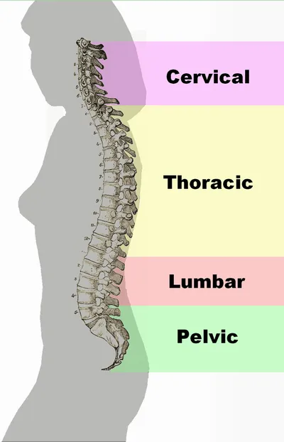
The adult spine has four alternating curves: cervical lordosis, thoracic kyphosis, lumbar lordosis, sacral kyphosis. The kyphoses are primary, present from fetal life; the lordoses are secondary and appear with postural development — the cervical one when the infant lifts its head, the lumbar one when it walks.
This alternation is not decorative. A straight column would transmit shocks without absorbing them; a curved column behaves like a spring, and its resistance is proportional to the square of the number of mobile curves, plus one: three curves multiply it by ten.
Each junction between two curves concentrates stress. This is where most injuries occur: the cervicothoracic, thoracolumbar and lumbosacral junctions.

The disc, and how it gives way
The intervertebral disc combines a central gelatinous nucleus, highly hydrated and incompressible, with a peripheral fibrous ring made of oblique, crisscrossing collagen lamellae. The nucleus spreads pressure in every direction; the ring contains it.
A herniated disc results from progressive tearing of the ring, through which the nucleus eventually bulges. By compressing a nerve root, it produces pain that follows that root's territory — sciatica for L5 or S1, femoral neuralgia for L3 or L4.
The adult disc is practically avascular and is nourished by diffusion from the vertebral endplates. It dries out with age, loses height and heals poorly. The most mobile levels — lower lumbar and lower cervical — are the most affected; the thoracic spine, held in place by the ribs, much less so.
Recognising a vertebra by its region
The spine has seven cervical, twelve thoracic and five lumbar vertebrae, then the sacrum, made of five fused vertebrae, and the coccyx, of three to five rudimentary pieces. Each region has its signature, which allows an isolated vertebra to be identified.
Cervical vertebrae have a small body, a transverse foramen in each transverse process, and a spinous process that is often bifid from C3 to C6. The lateral edges of their upper surface rise into hooks, the uncinate processes, which form uncovertebral joints with the vertebra above; osteoarthritis of these joints can narrow the intervertebral foramen and compress a nerve root.
Thoracic vertebrae carry costal facets on the body and on the transverse processes, for the heads and tubercles of the ribs. Their spinous process is long and slopes downwards, their articular facets face in an almost frontal plane. Lumbar vertebrae, finally, have the most massive body, no transverse foramen or costal facet, and articular facets oriented in a sagittal plane.
The ligaments and the vertebral canal
A system of ligaments binds the vertebrae together. The broad anterior longitudinal ligament runs along the front of the vertebral bodies and limits extension. The posterior longitudinal ligament runs inside the canal, along the back of the bodies; it narrows in the lumbar spine, leaving the posterolateral part of the disc unreinforced.
Posteriorly, the ligamenta flava unite the vertebral laminae. Rich in elastic fibres, hence their yellow colour, they form the posterior wall of the canal and help straighten the spine after flexion. The interspinous and supraspinous ligaments link the spinous processes.
The vertebral canal contains the spinal cord, surrounded by its meninges, and, outside the dura, the epidural space, filled with fat and venous plexuses: this is where the anaesthetic is injected in an epidural. The spinal nerves leave the canal through the intervertebral foramina, bounded by the pedicles of two neighbouring vertebrae, the disc and the zygapophyseal joint.
Movements of the spine
Each level allows only a few degrees, but together they give the spine great mobility. The orientation of the articular facets determines what is possible in each region.
The cervical spine is the most mobile in all directions. Its first two levels are specialised: the atlanto-occipital joint mainly provides flexion-extension of the head, the "yes", and the atlanto-axial joint provides rotation, the "no". The thoracic spine, held by the ribs and sternum, lends itself mainly to rotation. The lumbar spine allows wide flexion-extension and lateral bending, but very little rotation, because its sagittal facets interlock.
Trunk flexion does not happen only in the spine: beyond about sixty degrees, the pelvis tilts on the hips. Bending forward with straight legs works the hamstrings as much as the discs.
Deformities and injuries of the spine
Scoliosis is a three-dimensional deformity: a lateral curve, measured by the Cobb angle and considered significant beyond ten degrees, combined with rotation of the vertebrae. This rotation lifts the ribs on one side and creates a rib hump, visible when the subject bends forward. The most common form, idiopathic, appears in children and adolescents and worsens during growth.
In the sagittal plane, the curves can become exaggerated: thoracic hyperkyphosis, as in adolescent Scheuermann disease or in older people with osteoporosis, and lumbar hyperlordosis.
In older people, vertebral compression fractures occur after minimal trauma and mainly affect the thoracolumbar junction. In young adults, spinal fractures result from violent trauma; the major risk is then spinal cord injury, which requires immobilising the spine of any seriously injured person until it has been ruled out.
Spinal nerves and vertebral levels
There are thirty-one pairs of spinal nerves: eight cervical, twelve thoracic, five lumbar, five sacral and one coccygeal. There are eight cervical nerves for seven vertebrae: the first leaves between the occiput and the atlas, and the eighth between C7 and T1.
In the cervical spine, each nerve therefore leaves above the vertebra bearing its number; from the thoracic spine down, it leaves below. Since the cord ends at about L1-L2, the lumbar and sacral roots descend in the canal over several levels before reaching their foramen: they form the cauda equina.
A lumbar disc bulging backwards and outwards does not compress the root exiting at its level, which has already passed above in the foramen, but the one descending to the next level. Hence the classic correspondence: L4-L5 herniation, L5 root; L5-S1 herniation, S1 root.
58 answered questions go with this chapter in the practice section.
Sources and further reading
Reference textbooks
- Pierre Kamina, Anatomie clinique, Maloine.
- Frank H. Netter, Atlas d'anatomie humaine, Elsevier Masson.
Open access
- 7.3 The Vertebral Column — OpenStax, Anatomy and Physiology 2e.
Last revised : . Text licensed under CC BY-SA 4.0.