Finding your way around the body
Anatomical position, reference planes and directional terms.
The standard anatomical position
Every anatomical description assumes a conventional posture: subject standing, eyes looking straight ahead, upper limbs at the sides, palms facing forward, feet together. Without this convention, “in front” or “above” would have no stable meaning.
It is a convention, not a natural posture. Supination of the forearms, in particular, is not the resting position: it is chosen so that the radius and ulna lie parallel, which simplifies the description of their relations.
The three planes of space
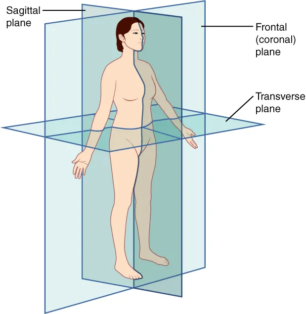
Three families of orthogonal planes divide the body and serve as the reference for every section, whether anatomical or radiological.
- The sagittal plane, vertical and anteroposterior, separates right from left. The median sagittal plane runs through the midline and defines two symmetrical halves.
- The frontal (or coronal) plane, vertical and side to side, separates front from back.
- The transverse (or horizontal, or axial) plane, perpendicular to the other two, separates top from bottom. It is the plane of the standard CT slice.

Directional terms
The pairs of terms are always used in opposition and always relative to the reference position.
- Superior / inferior: toward the head or toward the feet. Also called cranial and caudal.
- Anterior / posterior: toward the front or toward the back. Ventral and dorsal are their equivalents.
- Medial / lateral: toward the median sagittal plane or away from it.
- Proximal / distal: in the limbs, closer to or farther from the root of the limb.
- Superficial / deep: closer to or farther from the surface of the body.
Describing without ambiguity
Without it, terms become ambiguous. “In front” means nothing for a hand that can rotate; “anterior to the radius” only makes sense if you know where the radius is. The reference position makes every description reproducible.
Describing a movement: one plane, one axis
Every elementary movement takes place in a plane, around an axis perpendicular to it. Flexion and extension occur in the sagittal plane, around a transverse axis: flexing the elbow brings the forearm towards the arm, extending it moves them apart. Abduction and adduction occur in the frontal plane, around an anteroposterior axis: raising the arm out to the side is abduction of the shoulder.
Rotation occurs in the transverse plane, around a vertical axis. It is called medial when the front of the segment turns towards the midline, lateral in the opposite case. Circumduction is not an elementary movement: it chains flexion, abduction, extension and adduction, making the segment trace a cone.
Some joints have their own vocabulary. In the forearm, pronation turns the palm backwards and supination forwards. At the ankle, we speak of dorsiflexion and plantar flexion; in the foot, inversion turns the sole towards the midline and eversion outwards. At the thumb, opposition brings its pad against the pads of the other fingers.
- Sagittal plane, transverse axis: flexion and extension.
- Frontal plane, anteroposterior axis: abduction and adduction, lateral bending of the trunk.
- Transverse plane, vertical axis: medial and lateral rotation, pronation and supination.
Regions and cavities of the body
The body is divided into head, neck, trunk and limbs. The trunk comprises the thorax, the abdomen and the pelvis; the upper limb breaks down into shoulder, arm, elbow, forearm, wrist and hand, the lower limb into hip, thigh, knee, leg, ankle and foot. In anatomy, the arm refers only to the segment between shoulder and elbow, and the leg only to the segment between knee and ankle.
Organs are housed in cavities. Posteriorly, the cranial cavity continues as the vertebral canal: together they contain the central nervous system. Anteriorly, the diaphragm separates the thoracic cavity from the abdominopelvic cavity. The thoracic cavity contains two pleural cavities, one for each lung, and the mediastinum, which holds the heart in its pericardial sac.
Each visceral cavity is lined by a serous membrane — pleura, pericardium, peritoneum — made of two layers that slide over each other. This arrangement lets organs change volume and move, like the inflating lung or the contracting gut, without rubbing against the wall.
Surface anatomy: what can be found by hand
A few palpable landmarks locate deep organs and vertebral levels. The sternal angle, the ridge between the manubrium and the body of the sternum, lies at the level of the second rib and the disc between T4 and T5: this is the level of the tracheal bifurcation and of the boundary between superior and inferior mediastinum. The junction between the sternum and the xiphoid process corresponds to T9.
On the back, the C7 vertebra, called the vertebra prominens, has the most prominent spinous process of the neck. The root of the spine of the scapula corresponds to T3, its inferior angle to T7. Lower down, the line joining the tops of the iliac crests crosses the spine at about L4: this is the landmark for lumbar puncture.
On the abdomen, the umbilicus projects at about L3-L4. The transpyloric plane, halfway between the jugular notch of the sternum and the pubic symphysis, passes through L1: it crosses the pylorus, the neck of the pancreas, the renal hila and the origin of the superior mesenteric artery. McBurney's point, the projection of the base of the appendix, lies at the junction of the lateral third and medial two thirds of the line joining the right anterior superior iliac spine to the umbilicus.
Reading an imaging slice
An axial CT or MRI slice is viewed, by convention, as if standing at the feet of the patient lying down. The patient's right side therefore appears on the left of the screen, and the front of the body at the top. Forgetting this convention puts the liver on the left and the aorta on the wrong side of the spine.
CT measures X-ray attenuation, expressed in Hounsfield units: zero for water, about minus one thousand for air, around minus one hundred for fat, several hundred to over one thousand for compact bone. What attenuates strongly appears white, what attenuates little appears black. Iodinated contrast enhances vessels and well-perfused organs.
MRI does not measure density but the behaviour of protons in a magnetic field. On T1-weighted images, fat is bright and water is dark; on T2, water and fluids become white, which makes oedema and cerebrospinal fluid stand out. Ultrasound, finally, shows fluids as black and is stopped by air and bone, which reflect almost the whole beam.
A shared nomenclature for bodies that all differ
Anatomical names are set by an international nomenclature, the Terminologia Anatomica, published in 1998 and revised since. In French it did away with terms still common in everyday speech: fibula rather than péroné, ulna rather than cubitus, patella rather than rotule, scapula rather than omoplate. Eponyms, such as the Eustachian tube or the duct of Wirsung, are replaced by descriptive names — auditory tube, pancreatic duct.
This nomenclature describes a typical body, which does not exist. Anatomical variations are the rule: the course of an artery, the number of renal veins, the presence of an accessory muscle. They are harmless for the person, but a surgeon or radiologist unaware of them may injure what they did not expect to find.
Right-left asymmetry is also part of the normal plan: heart on the left, liver on the right, a three-lobed right lung. In situs inversus, which is rare, all organs are arranged as a mirror image, usually without any functional disorder.
61 answered questions go with this chapter in the practice section.
Sources and further reading
Reference textbooks
- Henri Rouvière, André Delmas, Anatomie humaine descriptive, topographique et fonctionnelle, Elsevier Masson.
- Pierre Kamina, Anatomie clinique, Maloine.
Open access
- 1.6 Anatomical Terminology — OpenStax, Anatomy and Physiology 2e.
Last revised : . Text licensed under CC BY-SA 4.0.