The lower limb
Pelvis, hip, knee, leg and foot.
An architecture built to bear weight
The whole design of the lower limb answers one constraint: transmitting body weight to the ground while allowing walking. Where the upper limb favors range, this one favors stability.
The hip illustrates this. Like the shoulder, it is a ball-and-socket joint with three degrees of freedom, but its femoral head sits deep in the acetabulum: it is far more stable and far less mobile.
The knee
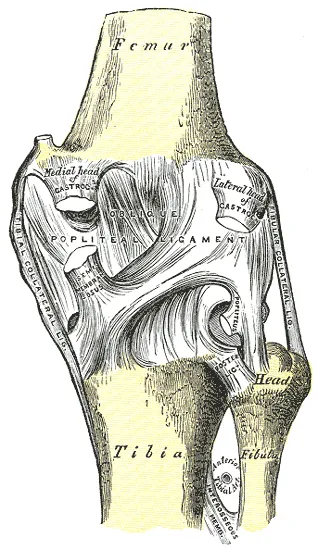
The knee combines a wide range of flexion-extension with demanding stability under load, despite poorly matched surfaces: convex femoral condyles on an almost flat tibial plateau.
Two systems compensate. The menisci, crescent-shaped fibrocartilages, improve congruence and spread the loads. The cruciate ligaments control forward-backward gliding, the collateral ligaments sideways gapping.

The foot
The tarsus has seven bones, including the talus, which receives the weight of the leg, and the calcaneus, which transmits it to the ground. Then come five metatarsals and fourteen phalanges — the big toe has only two, like the thumb.
The foot is not flat: it is organized as a vault, with two longitudinal arches and one transverse arch. This vault absorbs and returns energy with every step.
Carrying the body
The femoral neck makes an angle of about one hundred and twenty-five degrees with the shaft. This offset moves the shaft away from the pelvis and gives the hip abductors the lever arm that stabilizes the pelvis during walking, but it subjects the neck to considerable bending stress — hence its fragility as soon as spongy bone thins out.
A femoral neck fracture in older people combines three factors: this lever arm, osteoporosis and frequent falls. Its precarious blood supply also exposes to necrosis of the femoral head.
The lumbar and sacral plexuses
The lower limb is supplied by two plexuses. The lumbar plexus, formed by roots L1 to L4, gives the femoral nerve, which controls the quadriceps and supplies sensation to the front of the thigh, and the obturator nerve, which controls the adductors. The saphenous nerve, a sensory branch of the femoral, descends as far as the medial border of the foot.
The sacral plexus, formed by roots L4 to S3, gives the sciatic nerve, the largest nerve in the body. It leaves the pelvis below the piriformis muscle, descends on the back of the thigh, controls the hamstrings, then divides above the knee into the tibial nerve, for the posterior compartment of the leg and the sole, and the common fibular nerve, for the anterior and lateral compartments.
The superior and inferior gluteal nerves, from the same plexus, control the muscles of the buttock. The pudendal nerve supplies the perineum and the striated sphincters.
The compartments of the thigh and leg
The thigh has three compartments. The anterior contains the quadriceps, the knee extensor, and sartorius, supplied by the femoral nerve. The medial contains the adductors and gracilis, supplied by the obturator nerve. The posterior contains the hamstrings — semitendinosus, semimembranosus and biceps femoris — hip extensors and knee flexors, supplied by the sciatic nerve.
The leg also has three. The anterior contains tibialis anterior and the toe extensors, which lift the foot, supplied by the deep fibular nerve. The lateral contains the fibular muscles, evertors, supplied by the superficial fibular nerve. The posterior contains triceps surae — gastrocnemius and soleus, joined in the calcaneal tendon — and the deep flexors, supplied by the tibial nerve.
As in the forearm, these compartments are closed by poorly extensible fasciae. The anterior compartment of the leg is the most exposed to compartment syndrome, after a tibial fracture or intense exertion.
The femoral triangle and the vessels
Below the inguinal ligament, the femoral triangle is bounded by sartorius laterally and adductor longus medially. From lateral to medial lie the femoral nerve, the femoral artery and the femoral vein. The femoral pulse is felt halfway between the anterior superior iliac spine and the pubic symphysis; the vein, just medial to it, serves as a deep venous access.
Medial to the vein, a small space, the femoral canal, contains lymphatics. A loop of bowel can enter it: this is a femoral hernia, more common in women and more prone to strangulation than an inguinal hernia.
The femoral artery becomes the popliteal artery as it passes through the adductor hiatus, then divides below the knee into the anterior and posterior tibial arteries, the latter giving the fibular artery. The popliteal pulse is felt behind the knee, the posterior tibial behind the medial malleolus, the dorsalis pedis on the top of the foot.
The great saphenous vein begins in front of the medial malleolus and rises along the medial side of the limb to the groin, where it joins the femoral vein. The small saphenous vein passes behind the lateral malleolus and joins the popliteal vein. The deep veins, accompanying the arteries, are the site of deep vein thromboses, whose clot can travel to the lung.
Walking
A gait cycle runs from one heel strike to the next heel strike of the same foot. It includes a stance phase, about sixty percent of the cycle, and a swing phase, when the foot moves forward in the air. For short periods, both feet are on the ground: this is double support, which disappears when running.
During stance, the hip abductors — gluteus medius and minimus — keep the pelvis from dropping towards the swinging limb; the quadriceps cushions knee flexion on landing; triceps surae propels the body at the end of stance. During swing, the foot lifters, including tibialis anterior, keep the toes from catching the ground.
Each deficit has its limp. Weak gluteal muscles make the pelvis drop with every step; a paralysed tibialis anterior, from injury to the common fibular nerve at the neck of the fibula, forces the knee to be lifted high so the dropped foot can clear: this is the steppage gait. Hip pain shortens stance on the affected side: this is the antalgic limp.
61 answered questions go with this chapter in the practice section.
Sources and further reading
Reference textbooks
- Pierre Kamina, Anatomie clinique, Maloine.
- Frank H. Netter, Atlas d'anatomie humaine, Elsevier Masson.
Open access
- 8.4 Bones of the Lower Limb — OpenStax, Anatomy and Physiology 2e.
Last revised : . Text licensed under CC BY-SA 4.0.