Spinal cord, spinal nerves and cranial nerves
Segmental organization, roots, dermatomes and the twelve cranial nerve pairs.
The spinal cord
The spinal cord lies in the vertebral canal but in adults stops at about the L1-L2 level, while the canal continues down to the sacrum. This mismatch comes from growth: the spine lengthens more than the cord. Below its end, the canal only contains free roots, the cauda equina.
This mismatch is what allows lumbar puncture at L3-L4 or L4-L5, below the end of the cord: the needle pushes mobile roots aside instead of passing through nerve tissue.

Roots and spinal nerves
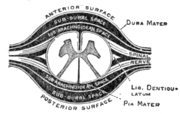
Each spinal nerve arises from two roots. The posterior root is sensory and carries the spinal ganglion, where the cell bodies lie; the anterior root is motor. They merge immediately, so the spinal nerve is mixed from its origin.
Each nerve supplies an area of skin, the dermatome, and a group of muscles, the myotome. Mapping these territories makes it possible to locate a lesion from the clinical examination alone.
The cranial nerves
Twelve pairs emerge from the brain. Some are purely sensory — olfactory, optic, vestibulocochlear; others purely motor — oculomotor, trochlear, abducens, accessory, hypoglossal; the rest are mixed — trigeminal, facial, glossopharyngeal, vagus.
The vagus nerve stands out by its territory: it leaves the head and neck to supply the thorax and much of the abdomen. It is the body's main parasympathetic nerve.
The cranial nerves and the autonomic system
The autonomic nervous system is divided into the sympathetic, whose fibers leave the thoracic and upper lumbar cord, and the parasympathetic, whose fibers leave through certain cranial nerves and the sacral cord. Their effects on a given organ are most often antagonistic.
The internal organisation of the spinal cord
In cross-section, the grey matter of the cord forms an H. The posterior horns receive sensory fibres; the anterior horns contain the motor neurons that command the muscles; between them, from T1 to L2, the lateral horns house the sympathetic neurons. The white matter surrounding the H is organised into posterior, lateral and anterior columns, carrying the major tracts.
Not all tracts cross the midline in the same place. The corticospinal tract, for voluntary movement, crosses at the lower medulla and descends in the lateral column on the side it controls. The posterior columns, which carry fine touch and position sense, ascend on the same side and cross only in the medulla. The spinothalamic pathway, which carries pain and temperature, crosses as soon as it enters the cord and ascends on the opposite side.
This layout explains Brown-Séquard syndrome, from hemisection of the cord: below the lesion, paralysis and loss of position sense on the same side, loss of pain and temperature sensation on the opposite side.
Dermatomes and myotomes: a few landmarks
Each root supplies a strip of skin, the dermatome, and a group of muscles, the myotome. A few landmarks are enough to locate a root or cord lesion from the examination.
- Upper limb, sensation: C6 at the thumb, C7 at the middle finger, C8 at the little finger.
- Upper limb, movement: C5 for shoulder abduction, C6 for elbow flexion, C7 for its extension, C8 for finger flexion, T1 for finger spreading.
- Trunk: T4 at the nipple, T10 at the umbilicus, L1 at the groin crease.
- Lower limb, sensation: L4 on the medial leg, L5 on the top of the foot and big toe, S1 on the lateral edge of the foot and the sole.
- Lower limb, movement: L2-L3 for hip flexion, L3-L4 for knee extension, L5 for lifting the big toe, S1 for plantar flexion.
Upper and lower motor neurons
Voluntary motor command passes through two neurons. The first, central or upper, starts in the motor cortex and descends in the corticospinal tract; the second, peripheral or lower, starts in the anterior horn of the cord or a brainstem nucleus and reaches the muscle through the nerve.
An upper motor neuron lesion produces a pyramidal syndrome: weakness, elastic increase in tone — spasticity —, brisk tendon reflexes and extension of the big toe when the sole is stroked, the Babinski sign. The muscle, still innervated, does not waste.
A lower motor neuron lesion, on the contrary, produces flaccid paralysis, with reduced tone, absent reflexes, muscle wasting and sometimes spontaneous twitches visible under the skin, fasciculations. The same logic applies to the face: a peripheral facial palsy affects the whole half of the face, forehead included, whereas a central lesion spares the forehead, which has bilateral control.
The twelve cranial nerves at a glance
Each cranial nerve has a precise role, which must be tested at the bedside.
- I, olfactory: smell. II, optic: vision.
- III, oculomotor: most eye muscles, eyelid elevation and pupil constriction. IV, trochlear: superior oblique muscle. VI, abducens: lateral rectus muscle.
- V, trigeminal: sensation of the face and muscles of mastication.
- VII, facial: muscles of facial expression, taste in the anterior two thirds of the tongue, tear and saliva secretion.
- VIII, vestibulocochlear: hearing and balance.
- IX, glossopharyngeal: taste in the posterior third of the tongue, sensation of the pharynx, parotid secretion.
- X, vagus: muscles of the pharynx and larynx, parasympathetic supply to the thoracic and abdominal viscera.
- XI, accessory: sternocleidomastoid and trapezius. XII, hypoglossal: muscles of the tongue.
The two autonomic systems
The sympathetic system arises from the lateral horns of T1 to L2: it is called thoracolumbar. Its short preganglionic neurons synapse in the chain of ganglia on either side of the spine or in prevertebral ganglia; the long postganglionic neurons most often release noradrenaline. The adrenal medulla, controlled directly by preganglionic fibres, is a glandular form of it.
The parasympathetic system is called craniosacral: it leaves through cranial nerves III, VII, IX and X, and through the sacral segments S2 to S4. Its long preganglionic fibres synapse in ganglia close to or within the walls of the organs; the short postganglionic fibres release acetylcholine.
A lesion of the sympathetic pathway to the head — in the neck, at the apex of the lung or in the brainstem — produces Horner syndrome: a slightly drooping upper eyelid, a constricted pupil and an eye that appears sunken, on the side of the lesion.
58 answered questions go with this chapter in the practice section.
Sources and further reading
Reference textbooks
- Pierre Kamina, Anatomie clinique, Maloine.
- Susan Standring (dir.), Gray's Anatomy. The Anatomical Basis of Clinical Practice, Elsevier.
Open access
- 13.4 The Peripheral Nervous System — OpenStax, Anatomy and Physiology 2e.
Last revised : . Text licensed under CC BY-SA 4.0.