Joints: classifying unions, describing movements
Synarthroses, amphiarthroses, diarthroses and degrees of freedom.
Classifying by mobility
The most useful classification rests on the range of movement allowed, itself dictated by the tissue that joins the bony parts.
- Synarthroses: fibrous union, almost no mobility. The sutures of the skull are the classic example.
- Amphiarthroses: cartilaginous union, limited but real mobility. The pubic symphysis and the intervertebral discs belong to this category.
- Diarthroses: synovial joints, mobile, with a joint cavity. These are joints in the everyday sense of the word.
The architecture of a diarthrosis
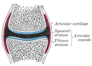
A synovial joint always combines the same elements: surfaces covered with hyaline cartilage, a fibrous capsule lined by a synovial membrane, and a cavity containing synovial fluid.
Depending on the joint, elements that improve congruence — the menisci of the knee, the labrum of the hip and shoulder — and ligaments that limit the range of motion are added.

Degrees of freedom
A diarthrosis is described by the number of axes around which it allows movement. A hinge joint such as the elbow has only one — flexion and extension. A condylar joint such as the wrist has two. A ball-and-socket joint such as the hip or shoulder has three, and also allows circumduction, which is not an elementary movement but a sequence linking flexion-extension with abduction-adduction.
Mobility versus stability
Every joint strikes a balance between two contradictory demands. The shoulder and the hip are both ball-and-socket joints with three degrees of freedom, but their compromises differ radically.
The glenoid cavity of the scapula is shallow and covers only a fraction of the humeral head: range is maximal, and stability rests almost entirely on the muscles and the labrum. This is why the glenohumeral joint is the most frequently dislocated joint in the body.
The acetabulum, by contrast, deeply encloses the femoral head, reinforced by powerful ligaments. The hip loses in range what it gains in stability: dislocating it takes violent trauma.
Why cartilage does not heal
Articular cartilage has neither blood vessels nor nerves. It is nourished by imbibition from the synovial fluid, helped by the pressure changes produced by movement: an immobilized joint starves.
This lack of vessels and nerves explains two major clinical facts. First, the absence of pain arising from cartilage: the pain of osteoarthritis comes from the subchondral bone, the synovium and the capsule, not from the cartilage itself. Second, the absence of repair: without vessels, no cells and no nutrients flow toward the lesion.
This is the central problem of joint trauma, and the reason why an extensive cartilage lesion tends to progress to osteoarthritis, sometimes all the way to joint replacement.
Fibrous and cartilaginous joints
Not all joints are synovial. In fibrous joints, bones are united by dense connective tissue, with no cavity. The sutures of the skull are the typical example; the distal tibiofibular syndesmosis unites two bones by a ligament and allows small movements; the gomphosis anchors the root of a tooth in its socket by the periodontal ligament.
In cartilaginous joints, the link is cartilage. Synchondroses are made of hyaline cartilage and are usually temporary: the growth plate is an example, ossifying at the end of growth. Symphyses combine fibrocartilage and remain for life: the pubic symphysis, the intervertebral discs.
These slightly mobile or immobile joints are not frozen. The pubic symphysis softens late in pregnancy under hormonal influence and widens the pelvis slightly; the cranial sutures, still open in infants, let the vault follow the growth of the brain.
Classifying synovial joints by shape
The shape of the articular surfaces dictates the number of degrees of freedom, that is, the number of axes around which movement is possible.
- Plane joints: nearly flat surfaces, simple gliding — joints between carpal bones, zygapophyseal joints.
- Hinge joints: a single transverse axis — humero-ulnar joint, interphalangeal joints.
- Pivot joints: a single longitudinal axis — proximal radio-ulnar joint, median atlanto-axial joint.
- Ellipsoid, or condylar, joints: two axes — radiocarpal joint, metacarpophalangeal joints.
- Saddle joints: two axes, surfaces concave one way and convex the other — the trapeziometacarpal joint of the thumb.
- Ball-and-socket joints: three axes — shoulder and hip.
What holds a joint together
Three elements contribute to stability. First the surfaces: a deeply seated head, like that of the femur in the acetabulum, resists dislocation by its shape alone. Then the ligaments, collagen bands that tighten at the end of the range and limit movement; they are passive brakes, not motors.
Finally the muscles, whose tone and reflex contraction press the surfaces together. At the poorly congruent shoulder, the rotator cuff plays this role constantly; at the knee, the quadriceps protects the joint when the ligaments are injured, hence the importance of muscle rehabilitation after a sprain.
Add to this fibrocartilaginous congruence elements — menisci, labrum, discs — and, in some joints, an intra-articular pressure below atmospheric pressure, which keeps the surfaces in contact.
Innervation, proprioception and injuries
The capsule and ligaments are rich in mechanoreceptors that constantly inform the nervous system about the position and movement of body segments: this is proprioception. A sprain damages these receptors along with the ligament fibres, which explains the feeling of instability and the risk of recurrence if proprioceptive rehabilitation is neglected.
According to Hilton's law, the nerves supplying the muscles that move a joint also supply that joint and often the overlying skin. This shared innervation explains referred pain: a hip disorder can present as knee pain, since the two joints partly share the territory of the obturator nerve.
A sprain is a ligament injury without loss of joint contact; a dislocation is a complete and lasting loss of that contact; a subluxation is a partial loss. A dislocation can injure nearby vessels and nerves, which must be examined before and after any reduction.
Osteoarthritis and arthritis
Osteoarthritis is a disease of the whole joint, starting with cartilage wear. It is accompanied by remodelling of the subchondral bone and the formation of osteophytes, bony outgrowths at the edges of the surfaces. On X-ray it shows as joint-space narrowing, subchondral sclerosis and osteophytes.
Its pain is called mechanical: worse with activity, relieved by rest, with brief morning stiffness. Arthritis, by contrast, is inflammation of the synovium — infectious, crystal-related such as gout, or autoimmune such as rheumatoid arthritis. Its pain is inflammatory: at night, with prolonged morning stiffness, often more than half an hour.
Aspirating the joint fluid settles the question. Mechanical fluid is clear and low in cells, fewer than two thousand per cubic millimetre; inflammatory fluid contains more. It is also examined for crystals and for bacteria.
60 answered questions go with this chapter in the practice section.
Sources and further reading
Reference textbooks
- Pierre Kamina, Anatomie clinique, Maloine.
- Henri Rouvière, André Delmas, Anatomie humaine descriptive, topographique et fonctionnelle, Elsevier Masson.
Open access
- 9.1 Classification of Joints — OpenStax, Anatomy and Physiology 2e.
- 9.4 Synovial Joints — OpenStax, Anatomy and Physiology 2e.
Last revised : . Text licensed under CC BY-SA 4.0.