The lungs
Lobes, bronchial segments, pleurae and the mechanics of breathing.
The bronchial tree and its asymmetry
The trachea divides at the carina into two main bronchi, which are not symmetrical. The right is wider, shorter and more vertical; the left is narrower, longer and more horizontal, since it must pass beneath the aortic arch.
This asymmetry has a classic consequence: an inhaled foreign body tends to enter the right bronchus, and aspiration pneumonias predominate on the right.
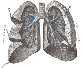
The lungs differ too. The right has three lobes separated by two fissures, the left only two, the heart taking the place of the third. Division continues into lobar and segmental bronchi, then bronchioles, down to the terminal and respiratory bronchioles.

The pleura, and what makes it indispensable
Two layers wrap each lung: the visceral pleura, adhering to its surface, and the parietal pleura, lining the chest wall. They are continuous with each other at the hilum.
Between them lies a potential cavity containing a few milliliters of fluid, and above all a negative pressure. It is this negative pressure that binds the lung to the chest wall: when the thorax widens, the lung follows, although no mechanical attachment connects them.
Air entering this cavity breaks the coupling. The lung, free to follow its elasticity, collapses: this is a pneumothorax. The same reasoning explains a pleural effusion, which compresses the lung instead of letting it expand.
Two circulations, two jobs
The lung receives two circulations. The pulmonary circulation, the functional one, brings the entire output of the right ventricle to the alveoli: the pulmonary trunk divides into arteries that follow the bronchi down to the alveolar capillaries. It is a low-pressure circuit — about fifteen millimetres of mercury on average — with low resistance.
The bronchial circulation, the nutritive one, arises from the thoracic aorta and supplies the bronchial walls. It takes no part in gas exchange, and some of its venous return drains into the pulmonary veins: this is one of the causes of the small physiological shunt.
At the alveolus, air and blood are separated by a barrier less than a micrometre thick: type I pneumocyte, fused basement membranes, endothelium. This thinness, spread over a surface of about seventy square metres, makes oxygen diffusion almost instantaneous.
Lobes, fissures and segments
The right lung is divided by two fissures into three lobes. The oblique fissure separates the lower lobe from the other two; the horizontal fissure separates the upper lobe from the middle lobe. The left lung has only an oblique fissure and two lobes; the anteroinferior tongue of its upper lobe, the lingula, is the equivalent of the right middle lobe.
Each lobe is subdivided into bronchopulmonary segments, ten on the right and eight to ten on the left depending on fusions. These segments have their own bronchus and artery, which makes them surgical units: one can be removed without compromising its neighbours.
The projection of the fissures onto the chest wall guides examination. The oblique fissure rises posteriorly to about the fourth rib and descends anteriorly along the sixth; the horizontal fissure follows the fourth costal cartilage on the right. As a result, the back of the chest mainly overlies the lower lobe, and the front the upper and middle lobes.
From bronchus to alveolus
The airway tree divides by successive branchings, about twenty in all. The first sixteen generations, from the trachea to the terminal bronchioles, form the conducting zone: they warm, humidify and filter the air, but take no part in gas exchange. Their volume, about one hundred and fifty millilitres in adults, is the anatomical dead space.
Beyond begins the respiratory zone: respiratory bronchioles, whose walls already bear a few alveoli, then alveolar ducts and sacs. The unit supplied by one terminal bronchiole forms the acinus, the functional unit of the lung. There are several hundred million alveoli.
The alveolar wall is lined by two cell types. Type I pneumocytes, very flat, cover most of the surface and form, with the capillary endothelium, the exchange barrier. Type II pneumocytes, cuboidal and fewer, secrete surfactant and act as stem cells to renew the epithelium. Alveolar macrophages patrol the surface and clear particles and germs.
The limits of the lungs and pleurae
In quiet expiration, the lower border of the lung crosses the sixth rib on the midclavicular line, the eighth on the midaxillary line and the tenth near the spine. The parietal pleura extends about two ribs lower: eighth, tenth and twelfth ribs. Between the two, a pleural space without lung, the costodiaphragmatic recess, fills during deep inspiration.
It is in this recess that a pleural effusion collects, blunting the costophrenic angle on an upright X-ray. It is also where it is aspirated, below the fluid level and just above the upper border of a rib.
Above, the pleural dome and the apex of the lung rise two to three centimetres above the clavicle, at the root of the neck. A subclavian vein puncture or a low neck wound can therefore injure the pleura and cause a pneumothorax.
Innervation and lymphatics
The bronchi receive autonomic innervation through the pulmonary plexus, located at the hilum. Vagal, parasympathetic fibres contract bronchial smooth muscle and stimulate mucus secretion. Bronchial relaxation depends mainly on circulating adrenaline, acting on beta-2 receptors: this is the target of asthma bronchodilators.
Lymph from the lung follows the bronchi to the hilar lymph nodes, then to those of the tracheal bifurcation and mediastinum. This route is followed by bronchial cancers and infections: enlarged hila on an X-ray often reflect enlarged nodes.
In smokers, alveolar macrophages loaded with particles and the lymphatics draining them stain the lung and hilar nodes black: this is anthracosis, visible to the naked eye at autopsy.
58 answered questions go with this chapter in the practice section.
Sources and further reading
Reference textbooks
- Pierre Kamina, Anatomie clinique, Maloine.
- Frank H. Netter, Atlas d'anatomie humaine, Elsevier Masson.
Open access
- 22.2 The Lungs — OpenStax, Anatomy and Physiology 2e.
Last revised : . Text licensed under CC BY-SA 4.0.