The kidneys and urinary tract
Position, cortex and medulla, the nephron and excretion.
Position and relations
The kidneys are retroperitoneal, lying against the posterior abdominal wall on either side of the spine. The right kidney sits slightly lower than the left, pushed down by the liver.
Each kidney is surrounded by a fibrous capsule, itself embedded in a fatty compartment that helps hold it in place and cushions shocks.
Cortex, medulla, nephron
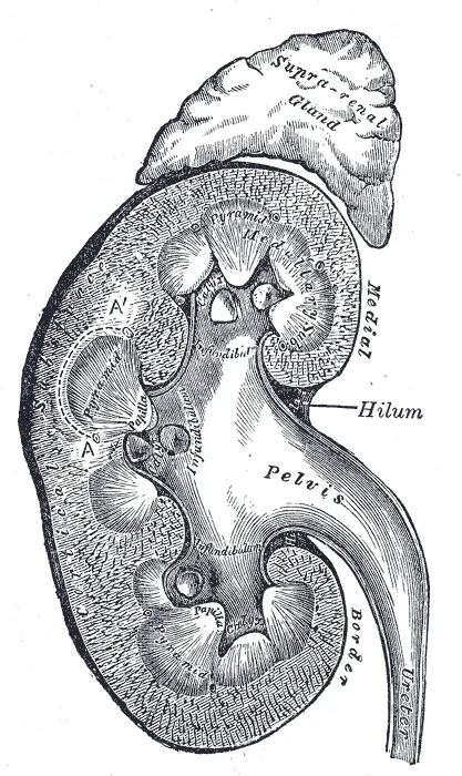
A frontal section shows a peripheral cortex and a medulla made of pyramids whose tips, the papillae, open into the calyces.
The functional unit is the nephron: a cortical renal corpuscle, where filtration takes place, extended by a tubule that dips more or less deeply into the medulla. It is this deep arrangement that allows urine to be concentrated.

The urinary tract
Urine flows into the calyces, the renal pelvis, then the ureter. The ureter is not a simple pipe: its smooth muscle wall propels urine by peristalsis, independently of gravity.
The ureter has three physiological narrowings — at the junction with the renal pelvis, where it crosses the iliac vessels, and where it passes through the bladder wall. These are where stones most often get stuck.
From corpuscle to calyx
The nephron begins with the renal corpuscle, located in the cortex: a tuft of capillaries, the glomerulus, enclosed in Bowman's capsule. Blood is filtered there under pressure, the barrier holding back cells and proteins.
The filtrate then runs through the proximal tubule, the loop of Henle, which dips into the medulla and comes back up, the distal tubule, and then the collecting duct. Each segment reabsorbs or secretes, so that the final urine has little in common with the initial filtrate.
The collecting ducts converge on the papillae, which open into the minor calyces, then the major calyces, the renal pelvis and finally the ureter.
An endocrine organ as much as an excretory one
The kidney does not just eliminate. It secretes renin, the starting point of the renin-angiotensin-aldosterone system that regulates blood pressure and blood volume. It produces erythropoietin, which stimulates red cell production. It completes the activation of vitamin D.
These three functions explain the consequences of chronic kidney disease far beyond the build-up of waste: hypertension, anemia, and bone disorders from a lack of active vitamin D.
The ureter, finally, does not carry urine by gravity but by peristalsis — which is what allows urine to reach the bladder even when lying down. When a stone suddenly blocks it, the urinary tract and capsule upstream are put under tension: this is the intense, paroxysmal pain of renal colic.
The coverings of the kidney
From inside out, the kidney is wrapped in a thin, poorly extensible fibrous capsule, then a layer of perirenal fat, itself contained in the renal fascia. Beyond the fascia, a second fatty layer, pararenal, separates it from the muscles of the posterior wall. The adrenal gland shares the renal space but occupies a separate compartment, so the kidney can be removed while leaving it in place.
The renal fascia forms a space closed above and laterally but open downwards along the ureter. This arrangement explains why a poorly supported kidney can drop, and why a collection of blood or pus around the kidney tracks downwards, towards the iliac fossa.
Perirenal fat protects and cushions the kidney. In a very thin person, its loss makes the kidney more mobile; on imaging, it forms a dark rim around the organ that helps detect infiltration.
The intrarenal circulation
The renal artery divides at the hilum into segmental arteries, which are end arteries, then into interlobar arteries that rise between the pyramids. At the junction of cortex and medulla they curve into arcuate arteries, from which interlobular arteries run towards the surface. These give off the afferent arterioles, one per glomerulus.
The kidney's peculiarity is to align two capillary networks in series. The first is the glomerulus, between the afferent and efferent arterioles: this is where filtration takes place. The second arises from the efferent arteriole: peritubular capillaries around the cortical tubules, and long straight vessels, the vasa recta, that dip into the medulla alongside the loops of Henle.
The medulla thus receives a small share of the flow, through these vasa recta whose hairpin course preserves the osmotic gradient. It lives permanently on the edge of hypoxia, which makes it vulnerable to falls in pressure and to certain drugs.
The bladder and the urethra
The bladder is a muscular reservoir in the lesser pelvis, behind the pubic symphysis, beneath the peritoneum, which covers only its upper surface. Empty, it stays hidden behind the pubis; full, it rises into the hypogastrium. The urge to urinate appears at about two to three hundred millilitres; normal capacity is about half a litre.
Its wall is made of smooth muscle, the detrusor, whose contraction empties the bladder. At the neck, an involuntary smooth sphincter maintains resting continence; lower down, around the urethra, the striated sphincter, voluntarily controlled through the pudendal nerve, allows urine to be held back when the urge becomes pressing.
The female urethra is short, three to four centimetres, and opens into the vestibule near the vagina: this is why lower urinary tract infections are much more common in women. The male urethra, sixteen to twenty centimetres long, passes through the prostate, the perineal floor, then the corpus spongiosum of the penis; it also carries semen.
Malformations and obstructions
The kidneys form in the pelvis and then ascend to the lumbar region. If their lower poles fuse, they form a horseshoe kidney, whose ascent is stopped by the inferior mesenteric artery that straddles it. The ureter can also be duplicated, partly or completely, which is often harmless.
Normally the ureter crosses the bladder wall obliquely: the filling bladder compresses this intramural course and prevents urine from flowing back. Too short a course allows vesicoureteric reflux, which exposes children to upper urinary infections, pyelonephritis.
An obstruction along the urinary tract — stone, compression, stricture — dilates the cavities above it: this is hydronephrosis. A stone migrating down the ureter causes renal colic, violent loin pain radiating forwards and downwards towards the iliac fossa and genitals, following the course of the ureter and the innervation it shares.
58 answered questions go with this chapter in the practice section.
Sources and further reading
Reference textbooks
- Pierre Kamina, Anatomie clinique, Maloine.
- Frank H. Netter, Atlas d'anatomie humaine, Elsevier Masson.
Open access
- 25.3 Gross Anatomy of the Kidney — OpenStax, Anatomy and Physiology 2e.
Last revised : . Text licensed under CC BY-SA 4.0.