The skull: neurocranium and face
Bones of the vault and base, sutures, cavities and passages.
Two sets, two functions
The skull divides into a neurocranium, the bony box that contains the brain, and a viscerocranium, or facial skeleton, which carries the sense organs and the entrance to the airway and digestive tract.
The neurocranium comprises a vault — frontal, parietals, squamous parts of the temporals, occipital — and a base, where the sphenoid holds the central position and articulates with all the other bones of the neurocranium.
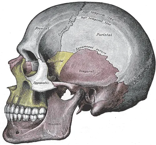

The sutures
The bones of the vault are joined by sutures, immobile fibrous joints. The coronal suture separates the frontal from the parietals, the sagittal suture joins the two parietals, and the lambdoid suture separates them from the occipital.
In the newborn, these sutures are not yet ossified and leave fontanelles between them. This flexibility lets the skull change shape during birth, then accommodates the rapid growth of the brain.
Vault and base, two regions, two risks
The skull divides into a vault and a base. The vault is made of flat bones — frontal, parietals, occipital, temporal squamae — joined by sutures. The base is a thick, irregular floor pierced by openings through which the cranial nerves and vessels pass.
Fractures do not mean the same thing in each. A fracture of the vault is often benign in itself, except over the temporoparietal squama, where the groove of the middle meningeal artery runs: its rupture causes an extradural hematoma, which compresses the brain within a few hours.
A fracture of the base can open a communication between the meningeal spaces and the air cavities of the face, hence cerebrospinal fluid leaking from the nose or ear and the risk of meningitis.
The newborn skull
At birth, the sutures are not ossified and leave membranous spaces between the bones, the fontanelles. The diamond-shaped anterior fontanelle closes around eighteen months; the triangular posterior one, around two months.
This flexibility has two roles. It lets the bones overlap as the head passes through the birth canal, reducing the head's diameter. It then accommodates the very rapid growth of the brain, which doubles in volume during the first year.
The anterior fontanelle is also a clinical window: bulging in raised intracranial pressure, sunken in dehydration, and accessible to transfontanellar ultrasound while it remains open.
The three levels of the skull base
Seen from above, the skull base descends in three steps, the cranial fossae. The anterior fossa, formed by the frontal bone, the cribriform plate of the ethmoid and the lesser wings of the sphenoid, supports the frontal lobes. The middle fossa, shaped like butterfly wings around the sella turcica, houses the temporal lobes. The posterior fossa, the deepest, formed mainly by the occipital bone and the back of the petrous temporal, contains the cerebellum and brainstem.
These fossae are pierced by openings through which the cranial nerves and vessels pass. Knowing their contents explains why a lesion at a single opening affects several nerves at once.
- Cribriform plate: filaments of the olfactory nerve (I).
- Optic canal: optic nerve (II) and ophthalmic artery.
- Superior orbital fissure: oculomotor (III), trochlear (IV), ophthalmic (V1) and abducens (VI) nerves, ophthalmic veins.
- Foramen rotundum: maxillary nerve (V2). Foramen ovale: mandibular nerve (V3). Foramen spinosum: middle meningeal artery.
- Internal acoustic meatus: facial (VII) and vestibulocochlear (VIII) nerves.
- Jugular foramen: glossopharyngeal (IX), vagus (X) and accessory (XI) nerves, and internal jugular vein.
- Hypoglossal canal: hypoglossal nerve (XII). Foramen magnum: medullospinal junction and vertebral arteries.
The facial skeleton and the sinuses
The face is built around the two maxillae, which carry the upper teeth, form the floor of the orbits and most of the palate. Added to them are the zygomatic bones, which form the cheekbones, the nasal, lacrimal and palatine bones, the inferior nasal conchae, the vomer and, the only mobile piece, the mandible.
The orbit is a pyramid whose wall is made of seven bones: frontal, sphenoid, ethmoid, lacrimal, maxilla, zygomatic and palatine. Its medial wall, formed mainly by the orbital plate of the ethmoid, is very thin, and its floor readily gives way when the eye takes a direct blow.
The paranasal sinuses are air cavities hollowed out in the bones around the nasal cavities: frontal, ethmoidal, maxillary and sphenoidal. They lighten the skull and give the voice its resonance. The maxillary sinus, the largest, drains into the middle meatus through an opening high on its medial wall, which hampers drainage in the upright position; the roots of the upper premolars and molars lie close to it, hence sinusitis of dental origin.
The mandible and chewing
The mandible has a horizontal body, carrying the lower teeth, and two vertical rami. Each ramus ends above in two projections: the condylar process, which articulates with the temporal bone, and the coronoid process, into which the temporalis muscle inserts. The inferior alveolar nerve, a branch of the mandibular nerve, runs through the mandibular canal and leaves by the mental foramen to supply the lower lip and chin.
Four muscles of mastication, all supplied by the mandibular nerve, move the mandible. Masseter and temporalis close the mouth with great force; the medial pterygoid assists them. The lateral pterygoid pulls the condyle and disc forwards: it protrudes the mandible and takes part in opening the mouth.
Head injuries
A fracture of the skull base often betrays itself by distant signs. Cerebrospinal fluid leaking from the nose, rhinorrhoea, indicates a breach of the anterior fossa, often at the cribriform plate; from the ear, otorrhoea, a fracture of the petrous temporal bone. Bruising around both eyes or behind the ear, over the mastoid, appears within hours. A dural breach exposes to meningitis.
In the vault, the region of the pterion, where the frontal, parietal, temporal bones and the greater wing of the sphenoid meet, is both thin and related to the middle meningeal artery. A fracture there can tear the artery and produce an extradural haematoma, a collection of blood between bone and dura, which progresses rapidly.
Fractures of the orbital floor, from a direct blow to the eye, can trap the inferior rectus muscle and limit upward gaze, with double vision. The infraorbital nerve, which runs in this floor, can be injured: the cheek and upper lip lose sensation.
60 answered questions go with this chapter in the practice section.
Sources and further reading
Reference textbooks
- Pierre Kamina, Anatomie clinique, Maloine.
- Henri Rouvière, André Delmas, Anatomie humaine descriptive, topographique et fonctionnelle, Elsevier Masson.
Open access
- 7.2 The Skull — OpenStax, Anatomy and Physiology 2e.
Last revised : . Text licensed under CC BY-SA 4.0.