The heart
Chambers, valves, coronary circulation and the cardiac cycle.
Four chambers, two pumps in series
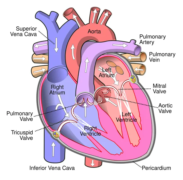
The heart is not one pump but two, side by side and working in series. The right heart receives deoxygenated blood from the venae cavae and propels it to the lungs through the pulmonary trunk. The left heart receives oxygenated blood from the pulmonary veins and ejects it into the aorta.

Four valves, one direction
The valves neither open nor close actively: they follow pressure gradients and impose a direction of flow.
- Tricuspid valve, between the right atrium and ventricle.
- Mitral valve, with two leaflets, between the left atrium and ventricle.
- Pulmonary valve, at the origin of the pulmonary trunk.
- Aortic valve, at the origin of the aorta.
Two pumps in series, at two pressure regimes
Connected in series, the two pumps must have the same output over time; otherwise blood would pile up on one side of the circuit.
What distinguishes them is not output but pressure. The pulmonary circulation is a low-pressure, low-resistance regime; the systemic circulation must perfuse the whole body, against a much higher resistance. Hence a left ventricular wall about three times thicker.
Shape follows: the left ventricle is conical, with a circular cross-section; the right is crescent-shaped, molded around it. This difference is the surest landmark for telling them apart on a section.
Feeding itself: the coronary circulation
The coronary arteries arise from the aorta just above the aortic valve, in the sinuses of Valsalva. The left coronary artery quickly divides into the anterior interventricular and circumflex arteries; the right runs in the right atrioventricular groove.
The left ventricular myocardium has a peculiarity: it is perfused mainly in diastole, because its own contraction squeezes the vessels running through it during systole. Any tachycardia shortens diastole first, and therefore reduces supply at the very moment demand increases.
Oxygen extraction there is already almost maximal at rest, unlike in other organs. The heart therefore cannot increase its extraction to cope with exertion: it has to increase coronary flow, which a stenosis prevents. This is the whole mechanism of exertional angina.
Position, surfaces and silhouette
The heart rests on the diaphragm, in the mediastinum, two thirds of it to the left of the midline. Its long axis runs obliquely downwards, forwards and to the left; its tip, or apex, formed by the left ventricle, usually projects into the fifth left intercostal space on the midclavicular line: this is where the apex beat is felt.
Its anterior, sternocostal surface is formed mainly by the right ventricle: this is what lies immediately behind the sternum. Its inferior, diaphragmatic surface belongs to both ventricles; its left, pulmonary surface to the left ventricle. The right border of the heart is formed by the right atrium.
On a frontal chest X-ray, the cardiac silhouette therefore has a right border formed by the right atrium and superior vena cava, and a left border made of four arcs from top to bottom: the aortic knob, the pulmonary trunk, the left auricle and the left ventricle. Enlargement of a chamber distorts the corresponding arc.
Inside the chambers
The right atrium receives both venae cavae and the coronary sinus. Its posterior part, where the venae cavae enter, is smooth; its anterior part and its auricle bear muscular ridges, the pectinate muscles. On the interatrial septum, the fossa ovalis is the remnant of the fetal foramen ovale.
The right ventricle, crescent-shaped around the left, has a wall covered with muscular trabeculae. One of them, the septomarginal trabecula, running from the septum to the base of a papillary muscle, carries the right bundle branch. Its smooth upper part forms the infundibulum, which leads to the pulmonary trunk.
The left atrium, the most posterior of the four chambers, receives the pulmonary veins; it touches the oesophagus, which allows it to be examined by transoesophageal echocardiography. Its narrow auricle is where clots form in atrial fibrillation. The left ventricle, thick-walled and circular in section, contains the mitral valve held by two papillary muscles.
The conduction system
The impulse arises in the sinus node, located in the wall of the right atrium near the entry of the superior vena cava. It spreads across both atria, then converges on the atrioventricular node, located in the interatrial septum above the opening of the coronary sinus.
Between atria and ventricles lies the fibrous skeleton of the heart, a set of connective-tissue rings that carry the valves and electrically insulate the two levels. The bundle of His is normally the only passage: it crosses this skeleton, then divides into two branches that descend on either side of the interventricular septum and ramify as the Purkinje network in the ventricular walls.
Ventricular contraction therefore begins at the septum and apex and progresses towards the base: blood is driven towards the arterial openings, which lie at the top. An accessory pathway bypassing the atrioventricular node, as in Wolff-Parkinson-White syndrome, removes the protective delay and exposes to tachycardias.
Innervation, pain and auscultation
The heart receives a dual autonomic supply. The sympathetic, from the upper thoracic segments, speeds up the rate and strengthens contraction; the parasympathetic, through the vagus nerve, slows the rate and atrioventricular conduction. Both mingle in the cardiac plexus, beneath the aortic arch.
Cardiac pain fibres travel back with the sympathetic to segments T1 to T4. The brain then attributes the pain to the skin territories of these segments: angina is felt behind the sternum and often radiates to the left arm, neck and jaw.
Auscultation is not done over the valves themselves, but where their sounds carry best, in the direction of flow. The aortic area is heard in the second right intercostal space at the edge of the sternum; the pulmonary area in the second left space; the tricuspid area at the lower left sternal border; the mitral area at the apex.
59 answered questions go with this chapter in the practice section.
Sources and further reading
Reference textbooks
- Pierre Kamina, Anatomie clinique, Maloine.
- Frank H. Netter, Atlas d'anatomie humaine, Elsevier Masson.
- Susan Standring (dir.), Gray's Anatomy. The Anatomical Basis of Clinical Practice, Elsevier.
Open access
- 19.1 Heart Anatomy — OpenStax, Anatomy and Physiology 2e.
Last revised : . Text licensed under CC BY-SA 4.0.