The upper limb
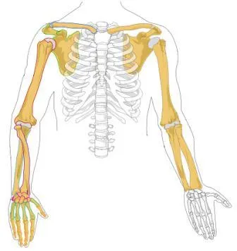
Shoulder girdle, arm, forearm and hand.
A girdle built for mobility
The shoulder girdle is attached to the trunk by a single bony joint: the sternoclavicular joint. The scapula, for its part, glides freely over the rib cage, held only by muscles.
This freedom explains the range of the shoulder, the most mobile joint in the body. It also explains its fragility: the glenoid cavity is shallow, and the glenohumeral joint is the one most often dislocated.

Elbow and pronation-supination
The elbow combines two functions within a single capsule. Flexion-extension occurs between the humerus on one side and the ulna and radius on the other, around a single axis.
Pronation-supination, by contrast, is a rotation of the radius around the ulna: the radius crosses the ulna in pronation and stays parallel to it in supination. This is why the standard anatomical position is described in supination.
The hand
The carpus has eight bones arranged in two rows. The metacarpus has five, and the digits fourteen phalanges — three per finger, except the thumb, which has only two.
The thumb owes its opposition to a saddle-shaped carpometacarpal joint, with two degrees of freedom, to which an automatic rotation of the metacarpal is added. This opposition is what makes the precision grip possible.
Three nerves, three recognizable deficits
The radial nerve runs in a groove on the back of the humerus and extends the wrist and fingers. A fracture of the humeral shaft can injure it: the hand drops and cannot be raised (wrist drop).
The median nerve passes through the carpal tunnel and notably provides opposition of the thumb. Its compression in this tunnel gives carpal tunnel syndrome, with tingling in the first three fingers, often at night.
The ulnar nerve passes behind the medial epicondyle, where it is superficial and exposed — it is the electric shock you feel when you bang your elbow (the “funny bone”). It controls most of the intrinsic muscles of the hand, hence the ulnar claw when it is damaged.
The brachial plexus
All the nerves of the upper limb arise from the brachial plexus, formed by the anterior rami of spinal nerves C5 to T1. These roots join into three trunks at the base of the neck, which divide and then regroup into three cords around the axillary artery: lateral, medial and posterior, named by their position relative to it.
The cords give off the major terminal nerves. The musculocutaneous nerve controls the flexors of the arm; the median nerve, most flexors of the forearm and the thenar muscles; the ulnar nerve, most intrinsic muscles of the hand; the axillary nerve, deltoid and teres minor; the radial nerve, all the extensors of the arm and forearm.
Violent traction on the limb or head can tear roots. Injury to the upper roots, C5 and C6, from a sudden downward pull on the shoulder, paralyses abduction and elbow flexion: the arm hangs in medial rotation, the hand turned backwards. Injury to the lower roots, C8 and T1, from upward traction on the arm, paralyses the intrinsic hand muscles, and the hand takes a clawed posture.
The muscle compartments of the arm and forearm
In the arm, a fibrous septum separates two compartments. The anterior compartment contains biceps, brachialis and coracobrachialis, supplied by the musculocutaneous nerve: these are the elbow flexors, biceps also being a powerful supinator. The posterior compartment contains triceps, the elbow extensor, supplied by the radial nerve.
In the forearm, the anterior compartment groups the flexors of the wrist and fingers and the pronators, arranged in superimposed layers. They are almost all supplied by the median nerve; only flexor carpi ulnaris and the medial half of flexor digitorum profundus are supplied by the ulnar nerve. The posterior compartment groups the extensors of the wrist and fingers and supinator, all supplied by the radial nerve.
This arrangement into compartments closed by poorly extensible fasciae has a consequence: swelling or bleeding within a compartment can raise its pressure enough to cut off the circulation. This is compartment syndrome, a surgical emergency requiring the fascia to be opened.
The shoulder in motion
Raising the arm does not move the glenohumeral joint alone. The scapula rotates on the thorax at the same time, turning the glenoid upwards: for every three degrees of arm elevation, about two occur at the glenohumeral joint and one through scapular rotation. This is the scapulohumeral rhythm.
Abduction is initiated by supraspinatus, then carried on by deltoid, while the other cuff muscles hold the humeral head in the glenoid. Beyond the horizontal, tilting of the scapula becomes essential; it is produced by trapezius and serratus anterior, which pull the inferior angle of the scapula outwards.
The supraspinatus tendon passes beneath the acromion, in a narrow space. Repeated friction during overhead movements gradually wears it: this is subacromial impingement, the leading cause of shoulder pain, which can end in a rotator cuff tear.
The vessels of the upper limb
The subclavian artery becomes the axillary artery at the lateral border of the first rib, then the brachial artery at the lower border of teres major. The brachial artery descends along the medial side of the arm with the median nerve and divides at the elbow crease into the radial and ulnar arteries, which rejoin in the palm through the superficial and deep palmar arches.
The pulse is taken at the radial artery, at the wrist, lateral to the tendon of flexor carpi radialis; blood pressure is measured by compressing the brachial artery in the arm. At the elbow crease, the brachial artery passes beneath an aponeurotic expansion of biceps, which separates it from the superficial veins.
The superficial venous network varies greatly, but two veins are constant: the cephalic vein on the lateral border of the limb, which drains into the axillary vein near the clavicle, and the basilic vein on the medial border. At the elbow crease a median vein links them: the usual site for blood samples.
Sensation in the hand and the carpal tunnel
The skin of the hand is shared by three nerves. On the palm, the median nerve supplies the thumb, index, middle finger and lateral half of the ring finger; the ulnar nerve, the medial half of the ring finger and the little finger. On the back, the radial nerve supplies the lateral part, the ulnar the medial part, the median reaching only the tips of the first three fingers. The most reliable area to test the radial nerve is the back of the first web space.
The carpal tunnel is an inextensible tunnel formed by the carpal bones behind and the flexor retinaculum in front. It contains the median nerve and nine tendons: four of flexor digitorum superficialis, four of flexor digitorum profundus and that of flexor pollicis longus. Anything that increases the volume of its contents — tenosynovitis, pregnancy, hypothyroidism — compresses the nerve.
The ulnar nerve passes outside the carpal tunnel, in the ulnar canal near the pisiform bone. This is why carpal tunnel syndrome spares the little finger.
58 answered questions go with this chapter in the practice section.
Sources and further reading
Reference textbooks
- Pierre Kamina, Anatomie clinique, Maloine.
- Frank H. Netter, Atlas d'anatomie humaine, Elsevier Masson.
Open access
- 8.2 Bones of the Upper Limb — OpenStax, Anatomy and Physiology 2e.
- 11.5 Muscles of the Pectoral Girdle and Upper Limbs — OpenStax, Anatomy and Physiology 2e.
Last revised : . Text licensed under CC BY-SA 4.0.