Bone tissue: architecture and remodeling
Compact and spongy bone, bone cells, growth and renewal.
Two architectures for a single material
Bone combines two arrangements of the same mineralized tissue. Compact bone, which is dense, forms the outer shell and the shaft of long bones: it withstands bending and twisting loads. Spongy bone, made of interlacing trabeculae, fills the epiphyses and the inside of short and flat bones.
This economy of material is no accident: the trabeculae of spongy bone line up along the lines of force running through the bone. A femoral neck shows bundles of trabeculae tracing the paths of compression and tension.
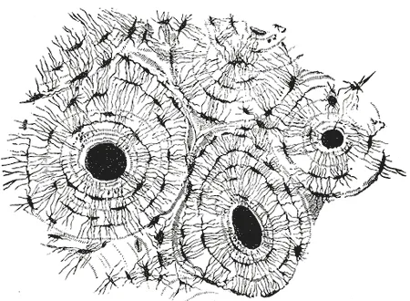

Three cell types, one balance
Bone tissue is alive and constantly renewed, through the action of three cell populations with complementary roles.
- Osteoblasts lay down the organic matrix, osteoid, which they then mineralize. Walled in by their own product, they become osteocytes.
- Osteocytes, housed in their lacunae and linked by fine processes, sense mechanical loads and direct remodeling.
- Osteoclasts, giant multinucleated cells, resorb the matrix by acidifying their digestion chamber.
A living bone, constantly rebuilding itself
Bone tissue is not an inert material laid down once and for all. It is continuously reworked: the osteoclast digs, the osteoblast fills, and the osteocyte, walled into its lacuna, senses loads and signals where reinforcement is needed.
This remodeling has three functions. It adapts the architecture to use — the trabeculae of spongy bone line up along the lines of force, and immobilization makes them disappear within a few weeks. It repairs microcracks before they spread. It makes bone a calcium reservoir that can be mobilized, under the control of parathyroid hormone.
The balance between resorption and formation shifts with age. Peak bone mass is reached around thirty; beyond that, resorption slowly wins out, and markedly faster in women after menopause, for lack of estrogens.
Growing in length, growing in width
A long bone grows by two distinct mechanisms, which must not be confused.
Growth in length happens at the growth plate, between the epiphysis and the metaphysis. The cartilage proliferates on one side and ossifies on the other, so that the bone lengthens without the plate moving. Its closure, at the end of puberty and driven by estrogens in both sexes, permanently ends growth in height.
Growth in width happens by periosteal apposition, at the surface, while the medullary cavity widens by internal resorption. The bone thus thickens without becoming massive — a tube resists bending better than a solid rod of the same mass.
Anatomy of a long bone
A long bone has a diaphysis, a tube of compact bone hollowed by a medullary cavity, and two swollen epiphyses, made of spongy bone under a thin cortex. Between the two, the metaphysis is the flared zone that, in children, borders the growth plate. The articular surfaces of the epiphyses are covered with hyaline cartilage.
Outside, the periosteum covers the whole bone except the articular surfaces. Its deep layer contains osteogenic cells, which provide growth in thickness and fracture healing. Inside, a thin cellular layer, the endosteum, lines the medullary cavity and the trabeculae.
Bone marrow fills the medullary cavity and the spaces of spongy bone. Red and blood-forming throughout the skeleton in children, it gradually turns into fatty yellow marrow in the diaphyses. In adults, blood formation persists mainly in the axial skeleton: vertebrae, sternum, ribs, hip bone, and the proximal ends of the femur and humerus.
A long bone receives a nutrient artery, which enters the diaphysis through an oblique foramen and supplies the marrow and the inner two thirds of the cortex. Metaphyseal, epiphyseal and periosteal vessels complete the supply. The periosteum is richly innervated; this is what makes fractures and bone bruises so painful.
Two ways to make a bone
Membranous, or intramembranous, ossification takes place directly in connective tissue: mesenchymal cells differentiate into osteoblasts and lay down matrix without a cartilage stage. It forms the bones of the skull vault, much of the face and the clavicle.
Endochondral ossification replaces a model of hyaline cartilage. A primary centre appears in the diaphysis around the seventh or eighth week of development; the secondary centres of the epiphyses mostly appear after birth. Between the two remains the growth plate, which allows growth in length until the end of puberty.
The growth plate is organised into successive layers, from epiphysis to diaphysis: a reserve zone, a proliferative zone where chondrocytes stack in columns, a hypertrophic zone, a calcification zone, then the ossification front where osteoblasts replace dead cartilage with bone.
What drives remodelling
The dialogue between osteoblasts and osteoclasts runs through a signalling system. Osteoblasts and osteocytes express RANKL, which binds the RANK receptor of osteoclast precursors and triggers their differentiation. They also secrete osteoprotegerin, a decoy that captures RANKL and restrains resorption. The ratio between the two sets the bone balance.
Hormones act largely through this lever. Parathyroid hormone increases RANKL and mobilises bone calcium; oestrogens restrain resorption, which explains the accelerated bone loss at menopause. Excess glucocorticoids inhibit osteoblasts: long-term corticosteroid therapy is the leading cause of secondary osteoporosis. Growth hormone and thyroid hormones take part in growth and turnover.
Osteoporosis treatments target these mechanisms. Bisphosphonates, bound to hydroxyapatite, are taken up by osteoclasts and inactivate them. Denosumab, an antibody against RANKL, blocks their differentiation. Other drugs, conversely, stimulate bone formation.
When bone becomes fragile
Osteoporosis combines reduced bone mass and deteriorated microarchitecture, which weaken bone. It is measured by bone densitometry: it is defined by a T-score of −2.5 or less, that is, a density two and a half standard deviations below that of a young adult. Its typical fractures affect the vertebrae, the femoral neck and the wrist, after minimal trauma.
Osteomalacia is a different disease: the matrix is produced normally but poorly mineralised, most often because of vitamin D deficiency. In children, whose growth plates are still open, the same deficiency causes rickets, with bone deformities and stunted growth.
A fractured bone heals through a haematoma, a soft fibrocartilaginous callus, a hard mineralised callus, then remodelling that restores the original architecture. Healing requires adequate immobilisation, a good blood supply and contact between fragments; otherwise it can fail and lead to non-union.
65 answered questions go with this chapter in the practice section.
Sources and further reading
Reference textbooks
- Pierre Kamina, Anatomie clinique, Maloine.
- Alan Stevens, James Lowe, Histologie humaine, Elsevier Masson.
Open access
- 6.3 Bone Structure — OpenStax, Anatomy and Physiology 2e.
- 6.4 Bone Formation and Development — OpenStax, Anatomy and Physiology 2e.
Last revised : . Text licensed under CC BY-SA 4.0.