The brain
Lobes, cortex, white matter, deep gray nuclei and blood supply.
A folded surface
The cerebral cortex is a sheet of gray matter a few millimeters thick. Its folding into gyri separated by sulci multiplies the available surface within a limited cranial volume: two-thirds of the cortex lies hidden in the depths of the sulci.
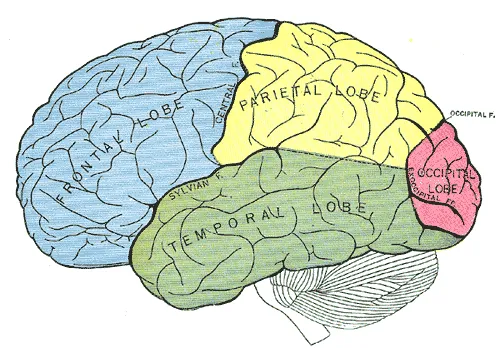
Two sulci serve as landmarks for dividing the lobes: the central sulcus separates the frontal lobe from the parietal lobe; the lateral sulcus sets off the temporal lobe.

Gray matter, white matter
Gray matter gathers the cell bodies of neurons. It forms the cortex, a peripheral sheet folded into gyri separated by sulci, and deep nuclei — the basal nuclei and thalamus.
The white matter beneath is made of myelinated axons. Three types of fibers make it up: association fibers link two regions of the same hemisphere; commissural fibers cross the midline, the corpus callosum being the largest; projection fibers link the cortex to underlying structures and to the spinal cord.
Blood supply and cerebrospinal fluid
The brain is supplied by two systems: the internal carotids at the front, the vertebral arteries at the back, which merge into the basilar trunk. They communicate at the base through the cerebral arterial circle, a backup arrangement — but it is complete in only a minority of people, which explains why the same occlusion does not have the same consequences from one person to another.
Cerebrospinal fluid is secreted by the choroid plexuses of the ventricles, flows toward the subarachnoid spaces and is reabsorbed by the arachnoid granulations. It provides mechanical protection, the brain floating and thus weighing only a fraction of its real weight.
An obstruction along this circuit produces hydrocephalus: fluid keeps being produced and builds up upstream.
The lobes and their functions
Each hemisphere is divided into lobes, named after the bones that cover them. The frontal lobe carries the primary motor cortex, in front of the central sulcus, the premotor areas and, further forward, the prefrontal cortex, which organises action, attention and social behaviour. In most people, Broca's area in the left frontal lobe controls speech production.
The parietal lobe receives bodily sensation in the postcentral gyrus and integrates space: a right parietal lesion can make the patient ignore the whole left half of space without noticing. The temporal lobe contains the auditory cortex, Wernicke's area, which allows language to be understood, and, on its medial surface, the hippocampus, essential for forming memories. The occipital lobe is devoted to vision; the primary visual cortex lines the calcarine sulcus.
Language is lateralised: the left hemisphere is dominant in about ninety-five percent of right-handers and a majority of left-handers. This is why a left middle cerebral artery lesion often combines right hemiplegia and aphasia.
Basal ganglia, diencephalon and brainstem
Deep in the hemispheres, the basal ganglia — caudate nucleus and putamen, which form the striatum, globus pallidus, subthalamic nucleus and substantia nigra — form loops with the cortex that regulate the initiation and scale of movements. Their dysfunction does not paralyse but disrupts movement: the slowness and rigidity of Parkinson disease, the involuntary movements of Huntington disease.
The diencephalon surrounds the third ventricle. The thalamus relays almost all sensory information to the cortex; the hypothalamus, below it, regulates temperature, hunger and thirst and controls the pituitary.
The brainstem links the brain to the spinal cord: midbrain, pons and medulla. All the major motor and sensory pathways pass through it; it contains the nuclei of cranial nerves III to XII and houses the centres that regulate breathing, heart rate and wakefulness. A small lesion there is therefore serious and often produces crossed syndromes, with a cranial nerve affected on one side and the body on the other.
The cerebellum
The cerebellum, housed in the posterior cranial fossa behind the brainstem, does not directly command muscles: it compares the intended movement with the movement performed and corrects continuously. It provides balance, coordination and precision of gestures, and regulates muscle tone.
Its midline part, the vermis, manages balance and trunk posture; the cerebellar hemispheres, limb coordination. Unlike the motor cortex, each cerebellar hemisphere acts on the side of the body on which it lies.
Cerebellar damage causes ataxia: a wide-based, drunken gait, movements that overshoot their target, inability to perform rapid alternating movements, a tremor that appears as the target is approached, and scanning speech. Alcohol, which particularly affects the vermis, produces a familiar temporary version.
The meninges and their spaces
Three membranes surround the brain. The thick dura mater adheres to the inner surface of the skull; its folds separate the two hemispheres — the falx cerebri — and the cerebrum from the cerebellum — the tentorium cerebelli —, and its splits form the dural venous sinuses. The thin arachnoid lines the dura; the pia mater follows the surface of the brain into the sulci.
Each space has its disease. Between bone and dura, the extradural space, normally only potential, fills when an artery is torn: extradural haematoma. Between dura and arachnoid, rupture of the veins crossing the space to reach the sinuses produces a subdural haematoma, common in older people and those on anticoagulants, sometimes after a minor knock. Beneath the arachnoid, the subarachnoid space contains cerebrospinal fluid and the arteries of the base: rupture of an aneurysm there causes subarachnoid haemorrhage, with a sudden, intense headache.
The dural folds partition the skull. A mass growing in one hemisphere can push the medial part of the temporal lobe under the free edge of the tentorium: this herniation compresses the oculomotor nerve, hence a dilated pupil on the same side, then the brainstem.
The ventricles and the arterial territories
Cerebrospinal fluid circulates through four ventricles. The two lateral ventricles, in the hemispheres, communicate through the interventricular foramina with the third ventricle, in the diencephalon. The cerebral aqueduct leads to the fourth ventricle, between the brainstem and cerebellum, which opens onto the subarachnoid spaces through one median and two lateral apertures.
Three arteries share each hemisphere. The anterior cerebral artery supplies the medial surface of the frontal and parietal lobes: its infarction mainly paralyses the opposite lower limb. The middle cerebral artery supplies the lateral surface: its infarction affects the opposite face and upper limb, and language if the dominant hemisphere is involved. The posterior cerebral artery supplies the occipital lobe: its infarction deprives the patient of the opposite half of the visual field, in both eyes.
This map makes the neurological examination a localising tool: the distribution of the deficit points to the artery involved before any imaging.
58 answered questions go with this chapter in the practice section.
Sources and further reading
Reference textbooks
- Pierre Kamina, Anatomie clinique, Maloine.
- Susan Standring (dir.), Gray's Anatomy. The Anatomical Basis of Clinical Practice, Elsevier.
Open access
- 13.2 The Central Nervous System — OpenStax, Anatomy and Physiology 2e.
- 13.3 Circulation and the Central Nervous System — OpenStax, Anatomy and Physiology 2e.
Last revised : . Text licensed under CC BY-SA 4.0.