Thoracic cage and trunk walls
Ribs, sternum, diaphragm and the mechanics of breathing.
A cage that must stay mobile
The rib cage protects the heart and lungs, but rigid protection would make breathing impossible. Its whole design is a compromise between strength and mobility.
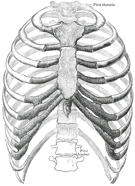
Twelve pairs of ribs articulate at the back with the thoracic vertebrae. At the front, the first seven join the sternum through their own cartilage, the eighth to tenth attach indirectly to the cartilage above, and the last two remain free — these are the floating ribs.

The diaphragm
The diaphragm is the main inspiratory muscle. Its contraction lowers its dome and increases the vertical diameter of the thorax: intrathoracic pressure falls and air flows in.
It is pierced by three major openings, at different levels: the inferior vena cava, the esophagus, then the aorta, from top to bottom and front to back.
The mechanics of breathing
Quiet expiration, on the other hand, is passive. The elastic recoil of the lung and chest wall is enough to push the air out, without muscular work. This point is often poorly remembered, yet it is central: a disease that hinders expiration, such as asthma, turns a free process into a costly effort.
During exertion, other muscles reinforce the action of the diaphragm. In inspiration, the external intercostals, already active at rest, are joined by the accessory muscles — scalenes, sternocleidomastoids — which raise the ribs; in expiration, the abdominal muscles and internal intercostals lower them.
The cage, and what it shelters
The intercostal neurovascular bundle runs beneath the lower edge of each rib, sheltered in a groove. This position dictates a procedure: any chest puncture is made at the upper edge of the rib below, to stay well away from the bundle.
The mediastinum fills the space between the two lungs. It contains the heart and its pericardium, the great vessels, the trachea and its bifurcation, the esophagus, the thymus in children, and the phrenic and vagus nerves.
Sternum, costal joints and rib movements
The sternum has three parts: the manubrium above, notched by the jugular notch and articulating with the clavicles and first ribs; the body, which receives the cartilages of ribs two to seven; and the xiphoid process below, cartilaginous for a long time. Its red marrow and superficial position make it a site for marrow aspiration.
Posteriorly, each rib articulates twice with the spine: its head with the vertebral bodies, its tubercle with the transverse process. Anteriorly, the costal cartilages articulate with the sternum. These joints let the rib pivot around an axis running through its neck.
The orientation of that axis changes the movement produced. The upper ribs rise forwards, like a pump handle, increasing the anteroposterior diameter of the thorax. The lower ribs swing outwards, like the handle of a bucket being lifted, increasing the transverse diameter. The diaphragm increases the vertical diameter.
The abdominal wall and the inguinal canal
In front, the two rectus abdominis muscles run from the thorax to the pubis, separated by the linea alba. Laterally, three flat muscles are layered: external oblique, internal oblique and transversus abdominis. Their aponeuroses form the rectus sheath; below the arcuate line, halfway between umbilicus and pubis, they all pass in front of the rectus, which then rests posteriorly only on the transversalis fascia.
This muscular belt flexes and rotates the trunk, but its main function is abdominal straining: contracting against a fixed diaphragm, it raises intra-abdominal pressure for coughing, defecation, urination or childbirth, and it supports the spine when lifting loads.
Above the inguinal ligament, the wall is crossed obliquely by the inguinal canal, about four centimetres long. It transmits the spermatic cord in men, the round ligament of the uterus in women, and the ilio-inguinal nerve. It opens deeply at the deep inguinal ring and superficially at the superficial ring.
The diaphragm in detail
The diaphragm is a muscular dome whose fibres converge on a central tendon. Posteriorly, it anchors to the lumbar spine by two crura: the longer right crus descends to L3, the left to L2. The right crus forms the muscular sling of the oesophageal hiatus, which contributes to the anti-reflux mechanism.
It is supplied by the phrenic nerves, from C3 to C5, which provide all its motor supply and the sensation of its central part. Irritation of this region — subphrenic abscess, blood in the peritoneum, gallbladder disease — can therefore be felt in the shoulder, whose skin belongs to the same spinal segment, C4. The periphery of the diaphragm receives its sensation from the intercostal nerves.
Two defects are classic. A hiatus hernia lets part of the stomach rise into the thorax through a widened oesophageal hiatus; it promotes reflux. A congenital diaphragmatic hernia, most often left posterolateral, lets abdominal viscera pass into the fetal thorax and hinders lung development.
The muscles of the back
The back muscles fall into three layers. The superficial layer, led by trapezius and latissimus dorsi, links the trunk to the upper limb: these are shoulder muscles that migrated onto the back, supplied by the accessory nerve for trapezius and by branches of the brachial plexus for the others.
The intermediate layer comprises the thin serratus posterior muscles, which attach to the ribs and play an accessory part in breathing. The deep layer is formed by the intrinsic back muscles: the erector spinae, in three columns, and the short transversospinalis muscles, running from one vertebra to another.
Only these deep muscles are supplied by the posterior rami of the spinal nerves. They are wrapped in the thoracolumbar fascia, a thick fibrous sheet that also gives attachment to latissimus dorsi and the flat abdominal muscles, and transmits forces between the upper limb, the trunk and the pelvis.
The breast
The mammary gland lies in the subcutaneous tissue of the anterior chest wall, in front of the fascia of pectoralis major, separated from it by a gliding space. It usually extends from the second to the sixth rib and continues towards the armpit as an axillary tail. The upper outer quadrant contains the most glandular tissue, and it is there that most cancers arise.
The gland is partitioned by suspensory ligaments, running from the deep fascia to the skin. A cancer invading these ligaments shortens them and pulls on the skin: retraction, dimpling, or a peau d'orange appearance when skin lymphatics are blocked.
Most of the lymph from the breast, about three quarters, drains to the axillary lymph nodes; the rest goes mainly to the parasternal nodes, along the internal thoracic vessels. This is why the search for nodal involvement starts in the armpit, where the first draining node, the sentinel node, is sought.
60 answered questions go with this chapter in the practice section.
Sources and further reading
Reference textbooks
- Pierre Kamina, Anatomie clinique, Maloine.
- Frank H. Netter, Atlas d'anatomie humaine, Elsevier Masson.
Open access
- 7.4 The Thoracic Cage — OpenStax, Anatomy and Physiology 2e.
- 22.3 The Process of Breathing — OpenStax, Anatomy and Physiology 2e.
Last revised : . Text licensed under CC BY-SA 4.0.