The liver and biliary tract
Position, dual blood supply, lobule and the path of bile.
A dual blood supply
The liver receives two blood supplies. The hepatic artery brings it oxygenated blood; the portal vein, which provides about three quarters of the flow, brings it the venous blood from the digestive tract, poor in oxygen but loaded with absorbed nutrients.
This arrangement makes the liver a mandatory filter: what the intestine absorbs reaches the systemic circulation only after passing through it — with the notable exception of fats, carried away by the lymph. This is the hepatic first pass, decisive in pharmacology.

Venous drainage
Blood from both sources mixes in the sinusoids, in contact with the hepatocytes, then returns to the inferior vena cava through the hepatic veins.
Bile, and the path of lipids
Bile is produced continuously by the hepatocytes. Between meals, with the sphincter of Oddi closed, it backs up into the gallbladder, which stores it and concentrates it by reabsorbing water.
The arrival of lipids in the duodenum triggers the release of cholecystokinin, which contracts the gallbladder and relaxes the sphincter. Bile then pours out, and its bile salts emulsify fats, multiplying the surface available to lipases.
Without bile, lipids and fat-soluble vitamins are hardly absorbed at all. A blockage of the bile ducts therefore produces pale, greasy stools, together with jaundice from accumulated bilirubin.
Shape, surfaces and attachments of the liver
The liver is the largest organ in the body: about one and a half kilograms in adults. It occupies the right hypochondrium and part of the epigastrium, under the dome of the diaphragm, protected by the ribs. It follows the diaphragm's movements, so its lower edge can be felt during deep inspiration.
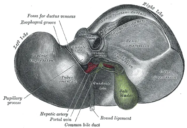
Its upper, diaphragmatic surface is smooth and convex. Its lower, visceral surface is marked by grooves forming an H: the crossbar is the hilum, or porta hepatis, where the portal vein and hepatic artery enter and the bile ducts leave; the vertical limbs house the gallbladder and the inferior vena cava on the right, the round ligament and the ligamentum venosum on the left. Besides the right and left lobes, these grooves outline the quadrate lobe in front and the caudate lobe behind.
The liver is attached to the diaphragm by the coronary ligament, extended by the triangular ligaments, and to the anterior wall by the falciform ligament. The free edge of the latter contains the round ligament, the remnant of the umbilical vein; the ligamentum venosum is the remnant of the fetal ductus venosus, which bypassed the liver.
The hepatic lobule
Under the microscope, the liver is made of hexagonal lobules. At the centre of each is a central vein; at the corners, portal tracts contain a triad: a branch of the portal vein, a branch of the hepatic artery and a bile duct. Between the two, plates of hepatocytes radiate, separated by widened, fenestrated capillaries, the sinusoids.
Blood from both sources mixes in the sinusoids and flows from the periphery to the central vein, then on to the hepatic veins. Bile, secreted by hepatocytes into tiny canaliculi, flows the opposite way, towards the bile ducts of the portal tracts.
The sinusoids are lined with resident macrophages, Kupffer cells, which clear portal blood of bacteria and debris from the gut. Between the endothelium and the hepatocytes, the space of Disse houses the stellate cells, which store vitamin A; activated by chronic injury, they produce collagen and drive fibrosis, then cirrhosis.
The bile ducts and gallstones
The bile ducts join into the right and left hepatic ducts, which form the common hepatic duct at the hilum. This receives the cystic duct from the gallbladder and becomes the common bile duct, which descends behind the head of the pancreas to open into the duodenum, together with the pancreatic duct, at the hepatopancreatic ampulla.
The gallbladder, lodged under the visceral surface of the liver, has a fundus, a body and a neck. It holds a few tens of millilitres of bile, which it concentrates by reabsorbing water; its fundus projects where the right costal margin meets the lateral border of the rectus abdominis.
Gallstones usually form in the gallbladder. A stone that briefly blocks the neck causes biliary colic; a prolonged, infected obstruction, cholecystitis. A stone that has migrated into the common bile duct blocks bile flow and causes jaundice; if it becomes infected, the result is cholangitis, which combines pain, fever and jaundice.
What the liver does
The liver sits at the crossroads of metabolism. It stores glucose as glycogen after meals and releases it between them; it makes glucose by gluconeogenesis during fasting. It synthesises albumin, the main plasma protein, and most clotting factors, including factors II, VII, IX and X, which depend on vitamin K. It assembles and exports lipoproteins.
It cleans the blood: it converts toxic ammonia into urea; it conjugates the bilirubin produced by red cell breakdown so it can be excreted in bile; it metabolises many drugs with its cytochromes. Finally, it stores iron, vitamin B12 and the fat-soluble vitamins.
Each lost function produces a sign of liver failure. Failure to conjugate or excrete bilirubin: jaundice. Failure to make albumin: oedema and ascites. Failure to make clotting factors: bleeding and a prolonged prothrombin time. Failure to clear ammonia: encephalopathy, altered consciousness.
Portal hypertension
In cirrhosis, fibrosis and regenerative nodules increase resistance to blood flow through the liver. Pressure rises in the portal vein and throughout the territory it drains: this is portal hypertension.
Blood then tries to reach the caval system through the portocaval anastomoses, normally insignificant, which dilate. At the lower oesophagus they form varices whose rupture causes severe gastrointestinal bleeding; around the umbilicus, a collateral venous circulation visible on the abdominal wall; in the rectum, venous dilatations.
Congestion also reaches the spleen, which enlarges and traps platelets, causing a low platelet count. Combined with low albumin, portal hypertension promotes the accumulation of fluid in the peritoneum, ascites.
58 answered questions go with this chapter in the practice section.
Sources and further reading
Reference textbooks
- Pierre Kamina, Anatomie clinique, Maloine.
- Frank H. Netter, Atlas d'anatomie humaine, Elsevier Masson.
Open access
- 23.6 Accessory Organs in Digestion: The Liver, Pancreas, and Gallbladder — OpenStax, Anatomy and Physiology 2e.
Last revised : . Text licensed under CC BY-SA 4.0.